. 2015 Jul;220(7):924-33.
doi: 10.1016/j.imbio.2015.01.013. Epub 2015 Feb 7.

The adult murine heart has a sparse, phagocytically active macrophage population that expands through monocyte recruitment and adopts an 'M2' phenotype in response to Th2 immunologic challenge

Affiliations

The adult murine heart has a sparse, phagocytically active macrophage population that expands through monocyte recruitment and adopts an 'M2' phenotype in response to Th2 immunologic challenge

Katie J Mylonas et al. Immunobiology. 2015 Jul.

Abstract

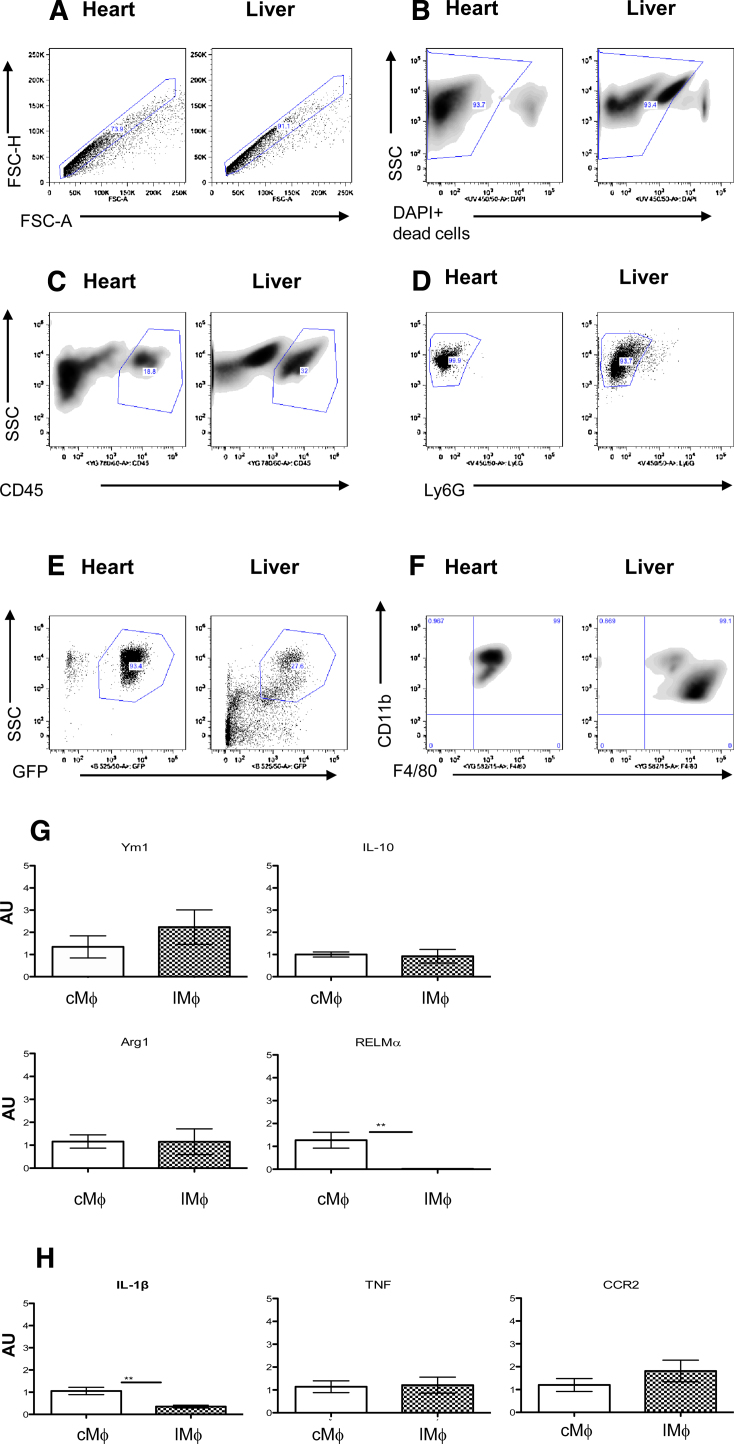
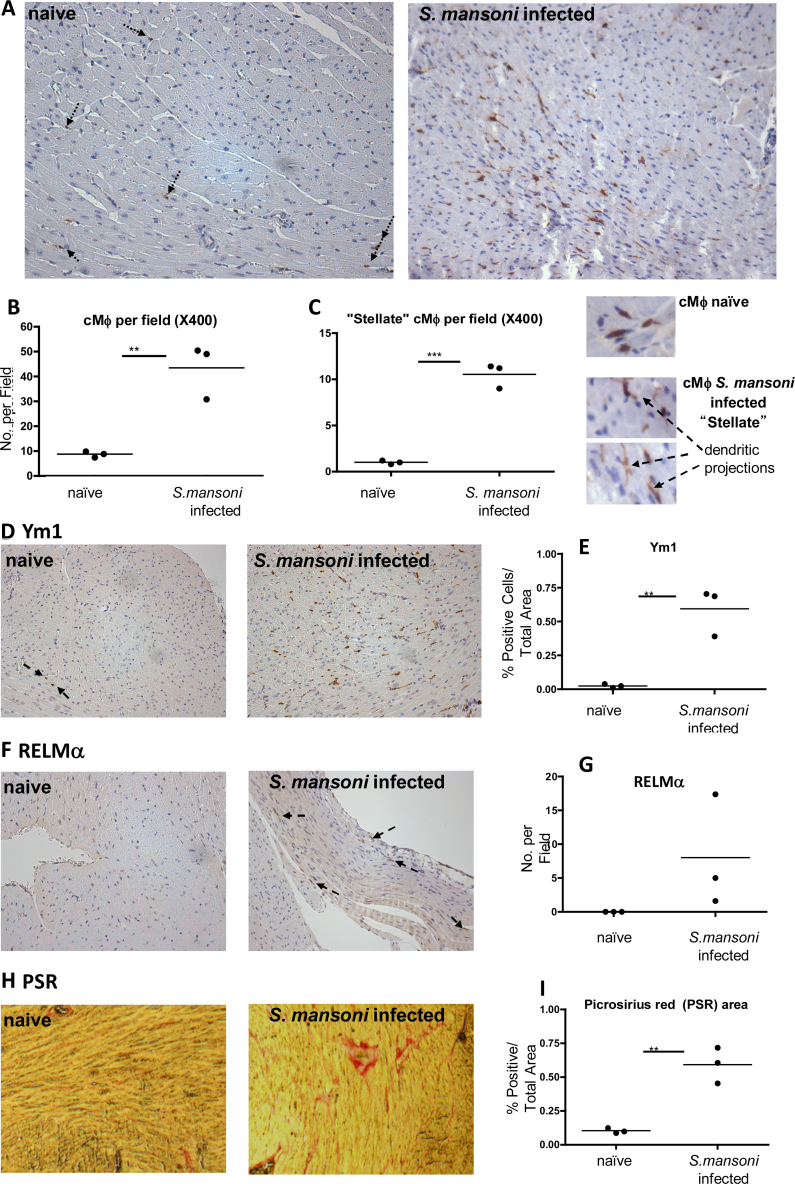
Tissue resident macrophages have vital homeostatic roles in many tissues but their roles are less well defined in the heart. The present study aimed to identify the density, polarisation status and distribution of macrophages in the healthy murine heart and to investigate their ability to respond to immune challenge. Histological analysis of hearts from CSF-1 receptor (csf1-GFP; MacGreen) and CX3CR1 (Cx3cr1(GFP/+)) reporter mice revealed a sparse population of GFP positive macrophages that were evenly distributed throughout the left and right ventricular free walls and septum. F4/80+CD11b+ cardiac macrophages, sorted from myocardial homogenates, were able to phagocytose fluorescent beads in vitro and expressed markers typical of both 'M1' (IL-1β, TNF and CCR2) and 'M2' activation (Ym1, Arg 1, RELMα and IL-10), suggesting no specific polarisation in healthy myocardium. Exposure to Th2 challenge by infection of mice with helminth parasites Schistosoma mansoni, or Heligmosomoides polygyrus, resulted in an increase in cardiac macrophage density, adoption of a stellate morphology and increased expression of Ym1, RELMα and CD206 (mannose receptor), indicative of 'M2' polarisation. This was dependent on recruitment of Ly6ChighCCR2+ monocytes and was accompanied by an increase in collagen content. In conclusion, in the healthy heart resident macrophages are relatively sparse and have a phagocytic role. Following Th2 challenge this population expands due to monocyte recruitment and adopts an 'M2' phenotype associated with increased tissue fibrosis.

Keywords: Heart; Helminth infection; Macrophage.


Figures

Fig. 1
Fig. 1
The adult murine heart contains resident CSF-1R+ and CX3CR1+ Mϕ that are evenly distributed throughout. (A) Representative micrograph of MacGreen CSF-1R GFP reporter mouse heart section (400× magnification). White arrows indicate representative GFP+ cells. (B) Cx3cr1GFP/+ heart section that has undergone anti-GFP immunohistochemistry (IHC) showing positive cells (black arrows indicate representative GFP stained cells; 400× magnification). Inlaid negative controls are shown at 200× magnification. Hearts sections from Cx3cr1GFP/+ mice were divided into the following subsections as outlined in the schematic transverse heart section (C). Left Ventricular Free Wall (LV FW; 1), Right Ventricular FW (RV FW; 2) and Septum (3), and subsections therein, i.e. subepicardial (outer) areas of LV and RV FWs (1.1 and 2.1; sub-epi), and subendcocardial (inner) areas of LV and RV FWs (1.2 and 2.2; sub-endo). The septum was divided into sub-LV-endocardial (3.1) and sub-RV-endocardial (3.2) sections, those being the areas closer to the LV and RV respectively (C). GFP+ cells were quantified in the LV, RV and septum (D) and the subsections of each (E). Representative micrographs showing perivascular areas (f; black arrows pointing to vessels, dashed arrows to Mϕ).
Fig. 2
Fig. 2
cMϕ are not overtly polarised. (A–F) Gating strategy for FACS of resident heart compared to resident liver Mϕ. Single cell suspensions from MacGreen hearts and livers were gated by granularity and size (by SSC, Side Scatter and FSC, forward scatter) and singlets (A) selected to exclude cell clumps (FSC- H/FSC-A; -height/-area). Dead cells (B) are excluded. CD45+ (C) and Ly6G negative cells are gated on to select leukocytes and to exclude neutrophils (D). Mϕ selected are GFP+ (CSF-1R+; E), CD11b+ and F4/80+ (F). (G, H) Realtime PCR analysis of cMϕ vs LMϕ. Comparison of M2-like Mϕ markers (G; Ym-1, IL-10, Arg1 and RELMα) and M1-like/inflammatory markers (H; iNOS, TNF and CCR2) by realtime PCR. in FACS sorted cMϕ (open columns) vs LMϕ (hashed columns, n = 6/7; *p < 0.05, ***p < 0.005; AU = arbitrary units, normalised to GAPDH).
Fig. 3
Fig. 3
Schistosoma mansoni infection changes the frequency, morphology and phenotype of heart resident Mϕ, and increases the collagen content of the heart. (A) Representative Cx3cr1GFP/+ heart sections from naïve and S. mansoni infected mice that have undergone anti-GFP IHC showing positive cells (black arrows indicate representative GFP stained cells in naïve mouse hearts. GFP positive cells are clear in infected section; 200× magnification). Number of cMϕ (B) and “stellate” Mϕ (C left) per field in naïve vs S. mansoni infected mice. Representative micrographs comparing stellate Mϕ (with dendritic projections) vs “typical” cMϕ (C right). (D) Representative heart sections from naïve and S. mansoni infected mice that have undergone Ym1 IHC showing positive cells (black arrows indicate representative GFP staining in naïve hearts; 200× magnification). (E) Quantification of Ym1+ cells per area in naïve vs S. mansoni infected heart sections. (F) RELMα+ cells in naïve (left) vs S. mansoni infected (right) mouse heart sections (black arrows indicate representative GFP stained cells; 200× magnification). (G) Quantification of RELMα staining in naïve vs S. mansoni infected heart sections (statistical analysis could not be carried out as no RELMα was detectable in naïve hearts). (H) Picrosirius Red staining indicating collagen content in naïve vs S. mansoni infected heart sections and quantification of % positive area (I). **p < 0.01, ***p < 0.005.
Fig. 4
Fig. 4
The presence of a H. polygyrus infection increases the frequency of heart resident Mϕ and this is largely dependent on monocyte recruitment. (A) Representative micrograph of GFP IHC in heart section from naïve and infected Cx3cr1GFP/+ mice. (B) Number of cMϕ (GFP+) and “stellate” cMϕ (C) per field in Cx3cr1GFP/+ in naïve (open columns) vs mice that had been infected for 28 days with the GI helminth H. polygyrus (filled columns). (D) Representative micrograph of Ym1 staining of a d28 infected heart (left panel) and quantification of Ym1+ cells per area in naïve vs 28 days post-infected mice (right panel). (E) Representative histograms of CD45+ GFP+ cells in the hearts of naïve and infected mice (left panel; dotted lines represent GFP-control), with quantification of positive cells acquired by flow cytometry (right). (F) Representative scatter profiles of mannose receptor (CD206) expressing cMϕ (left and middle panel) and positive cell numbers (right panel) in naïve vs d28 H. polygyrus infected hearts quantified by flow cytometry. (G) Quantification of Ly6Chigh (CCR2+) monocytes in the blood of WT naïve (open column), CCR2KO naïve (grey and white striped column) mice, and WT (black filled column) and CCR2KO (grey filled column) animals that have been infected for 28 days with H. polygyrus. (H) Quantification of cMϕ (total Mϕ-left, Ly6Clow-middle and Ly6Chigh-right) acquired by flow cytometry in naïve WT and CCR2KO mice or those that have been infected with H. polygyrus. *p < 0.05, **p < 0.01, ***p < 0.005.

References

    1. Allen J.E., Wynn T.A. Evolution of Th2 immunity: a rapid repair response to tissue destructive pathogens. PLOS Pathogens. 2011;7(5):e1002003. - PMC - PubMed
    1. Azzawi M., Hasleton P.S., Kan S.W., Hillier V.F., Quigley A., Hutchinson I.V. Distribution of myocardial macrophages in the normal human heart. J. Anat. 1997;191:417–423. - PMC - PubMed
    1. Boring L., Gosling J., Chensue S.W., Kunkel S.L., Farese R.V., Jr., Broxmeyer H.E., Charo I.F. Impaired monocyte migration and reduced type 1 (Th1) cytokine responses in C–C chemokine receptor 2 knockout mice. J. Clin. Investig. 1997;15:2552–2561. - PMC - PubMed
    1. Davies L.C., Jenkins S.J., Allen J.E., Taylor P.R. Tissue-resident macrophages. Nat. Immunol. 2013;14:986–995. - PMC - PubMed
    1. Epelman S., Lavine K.J., Beaudin A.E., Sojka D.K., Carrero J.A., Calderon B., Brija T., Gautier E.L., Ivanov S., Satpathy A.T., Schilling J.D., Schwendener R., Sergin I., Razani B., Forsberg E.C., Yokoyama W.M., Unanue E.R., Colonna M., Randolph G.J., Mann D.L. Embryonic and adult-derived resident cardiac macrophages are maintained through distinct mechanisms at steady state and during inflammation. Immunity. 2014;40:91–104. - PMC - PubMed

Publication types

MeSH terms