Review
. 2015 Aug;25(6):487-96.
doi: 10.1016/j.tcm.2015.01.005. Epub 2015 Jan 16.

Ionic mechanisms of arrhythmogenesis

Affiliations
Review

Ionic mechanisms of arrhythmogenesis

Justus M Anumonwo et al. Trends Cardiovasc Med. 2015 Aug.

Abstract

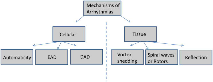
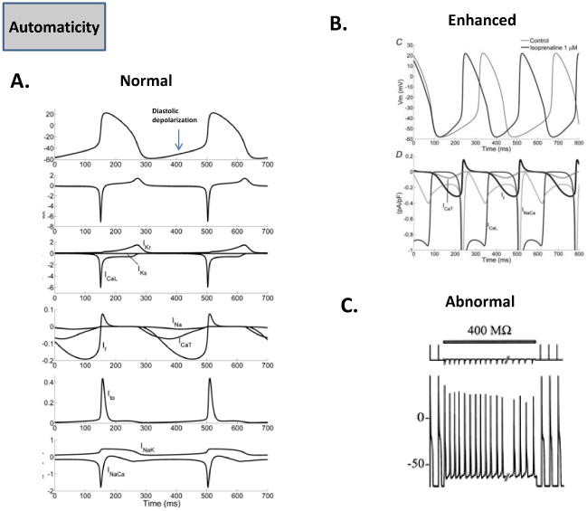
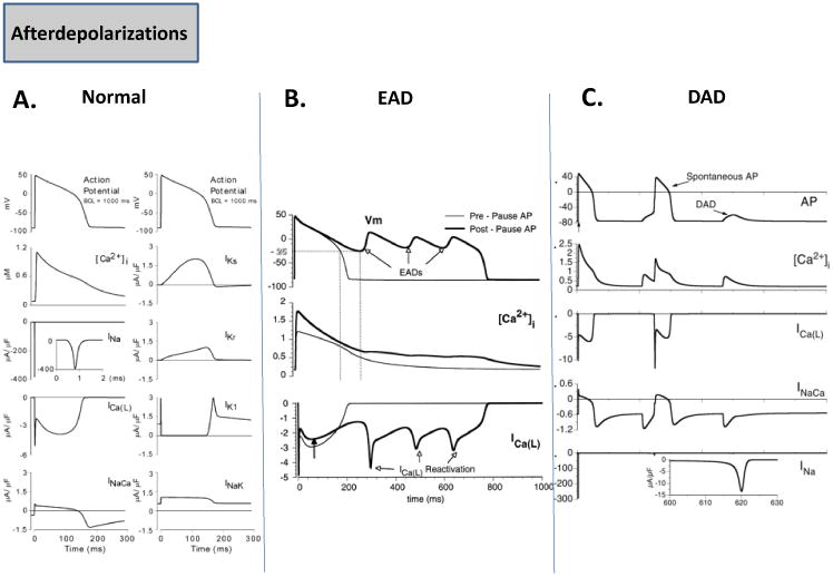
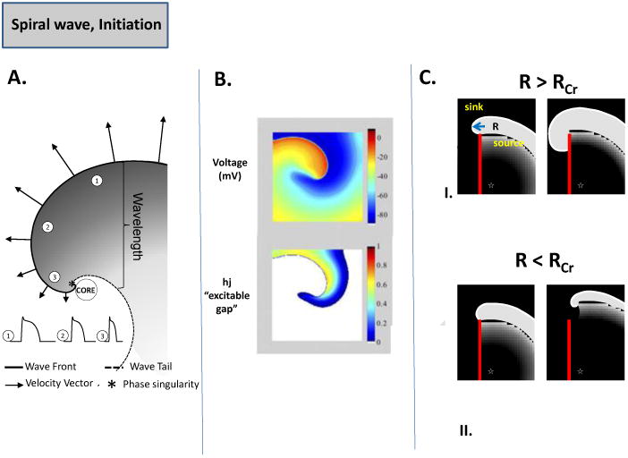
The understanding of ionic mechanisms underlying cardiac rhythm disturbances (arrhythmias) is an issue of significance in the medical science community. Several advances in molecular, cellular, and optical techniques in the past few decades have substantially increased our knowledge of ionic mechanisms that are thought to underlie arrhythmias. The application of these techniques in the study of ion channel biophysics and regulatory properties has provided a wealth of information, with some important therapeutic implications for dealing with the disease. In this review, we briefly consider the cellular and tissue manifestations of a number of cardiac rhythm disturbances, while focusing on our current understanding of the ionic current mechanisms that have been implicated in such rhythm disturbances.


Conflict of interest statement

Conflicts of Interest: JM Anumonwo: None, SV Pandit: None

Figures

Fig. 1
Fig. 1. Mechanisms of Arrhythmias
The various cellular and tissue mechanisms of arrhythmias are depicted.
Fig. 2
Fig. 2. Automaticity
(A.) The normal SAN pacemaker activity and the main underlying ionic currents are shown [8]. (B.) Enhanced automaticity in the SAN pacemaker during adrenergic stimulation, and underlying ionic determinants are shown [8]. (C.) Shows coupling of Purkinje cell to a ventricular cell model via a resistance of 400 MΩ. The ventricular cell is clamped at Vrest of -50 mV, which induces abnormal automaticity in the Purkinje cell [13].
Fig. 3
Fig. 3. Afterdepolarizations
(A.) The normal ventricular action potential and underlying ionic currents are depicted [17]. (B.) Representative simulation of an action potential in LQT2 at a basic cycle length of 500 ms (pre-pause AP), and an EAD occurring after a pause of 1500 ms is shown. Also shown is the underlying Ca2+ transient as well as the repetitive reactivation of ICaL [19]. (C.) Representative simulation of a DAD and a spontaneous action potential during Ca2+ overload, and the underlying Ca2+ transient, ionic currents (ICaL, INaCa, INa), are shown [25].
Fig. 4
Fig. 4. Spiral Waves, and their Initiation
(A.) Schematic of the spiral wave: Electrotonic effects of the core decrease conduction velocity (arrows), action potential duration (representative examples shown from positions 1, 2 and 3) and wavelength (the distance from the wave front (black line) to the wave tail (dashed line) [7]. As CV decreases and wavefront curvature becomes more pronounced, a point singularity occurs as the wave front and tail meet “*” [34]. (B.) Computer simulation of spiral wave. Top panel: snapshot of the transmembrane voltage distribution during simulated reentry in chronic AF conditions in a 2D sheet incorporating human atrial ionic math models [35]. Bottom panel: snapshot of inactivation variables of sodium current, “h.j” during reentry. (C.) Schematic of spiral wave initiation. As a wave progresses along an obstacle (red line) the curvature of the wave at the edge of the obstacle will determine if the wave detaches. If the curvature of the wavefront (R) at the edge of the obstacle is greater than the critical curvature for detachment (RCr) the wave remains attached; if R is less than RCr, when excitability is reduced (due to reduced INa, increased IK1, or enhance fibrosis) the wave will detach from the obstacle and initiate reentry [7].
Fig. 5
Fig. 5. Maintenance of Spiral Waves
(A.) Spirals in wild-type (WT) mouse hearts and transgenic (TG) mice where IK1 was overexpressed [43]. (B.) Spirals two-dimensional layers of rat neonatal ventricular myocytes in control, and where IKs was overexpressed via adenovirus [45]. (C.) Spirals in two-dimensional layers of rat neonatal ventricular myocytes in control, and where IKr was overexpressed via adenovirus [46].
Fig. 6
Fig. 6. Reflection
(A.) Shows transmembrane potentials in proximal (P) and distal (D) canine false tendons, with reflection giving rise to a second action potential in the P region [55]. (B.) The top left panel shows two rectangular patches of NRVM connected via a narrow isthmus. The bottom left panel shows time-space plot (Y axis, time, X axis-space) of impulse propagation (green) along the dotted line shown in the above panel. The right panel shows time plot of action potentials along the green line [56].

Comment in

References

    1. Rubart M, Zipes DP. Mechanisms of sudden cardiac death. J Clin Invest. 2005 - PMC - PubMed
    1. Heijman J, Voigt N, Nattel S, Dobrev D. Cellular and molecular electrophysiology of atrial fibrillation initiation, maintenance, and progression. Circ Res. 2014 Apr 25;114(9):1483–99. - PubMed
    1. Cerrone M, Cummings S, Alansari T, Priori SG. A clinical approach to inherited arrhythmias. Circ Cardiovasc Genet. 2012 Oct 1;5(5):581–90. Review. - PubMed
    1. Tomaselli GF, Zipes DP. What causes sudden death in heart failure? Circ Res. 2004 Oct 15;95(8):754–63. Review. - PubMed
    1. Behr ER, Roden D. Drug-induced arrhythmia: pharmacogenomic prescribing? EurHeart J. 2013 Jan;34(2):89–95. Review. - PMC - PubMed

Publication types