Review
. 2015 Mar:42:11-55.
doi: 10.1016/j.matbio.2015.02.003. Epub 2015 Feb 18.

Proteoglycan form and function: A comprehensive nomenclature of proteoglycans

Affiliations
Review

Proteoglycan form and function: A comprehensive nomenclature of proteoglycans

Renato V Iozzo et al. Matrix Biol. 2015 Mar.

Abstract

We provide a comprehensive classification of the proteoglycan gene families and respective protein cores. This updated nomenclature is based on three criteria: Cellular and subcellular location, overall gene/protein homology, and the utilization of specific protein modules within their respective protein cores. These three signatures were utilized to design four major classes of proteoglycans with distinct forms and functions: the intracellular, cell-surface, pericellular and extracellular proteoglycans. The proposed nomenclature encompasses forty-three distinct proteoglycan-encoding genes and many alternatively-spliced variants. The biological functions of these four proteoglycan families are critically assessed in development, cancer and angiogenesis, and in various acquired and genetic diseases where their expression is aberrant.

Keywords: Angiogenesis; Cancer growth; Glycosaminoglycan; Growth factor modulation; Proteoglycan.


Figures

Fig. 1
Fig. 1
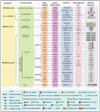
A comprehensive classification of proteoglycans. The four families are based on their cellular and subcellular location, homology at the protein and genomic levels and the presence of unique protein modules which are often shared by members of a given class. The key for the various modules is provided in the bottom panel. For additional details about structure and function, please consult the text.
Fig. 2
Fig. 2
Schematic representation of the cell surface proteoglycans, which comprise transmembrane type I (the N-terminus is outside of the plasma membrane) proteoglycans (four syndecans, CSPG4/NG2, betaglycan and phosphacan) and six GPI-anchored proteoglycans, glypicans 1–6. The type of GAG chain and the major protease sensitive sites are indicated. The key for the various modules is provided in the bottom panel.
Fig. 3
Fig. 3
Schematic representation of the pericellular proteoglycans, which comprise perlecan agrin, and collagens XVIII and XV. The collagenous (COL) and non-collagenous (NC) domains of collagen XVIII are numbered on the top and bottom of the lower schematics. For brevity only the structure of collagen XVIII is shown. The key for the various modules is provided in the bottom panel.
Fig. 4
Fig. 4
Schematic representation of the hyaluronan- and lectin-binding proteoglycans (hyalectans), which comprise aggrecan, versican, neurocan and brevican. The full-length versican (V0) and the three splice variants lacking GAGα (V1), GAGβ (V2) or both GAGα and GAGβ (V3) are shown. A new variant, V4, containing a portion of GAGβ is not shown. A GPI-anchored form of brevican is also not shown in the graphic. The dotted circles specify the globular domains (G1–G3) shared by the other hyalectans. These modules are compose of ~100 amino acids and have a characteristic consensus sequence with four disulfide-bonded Cys residues. The key for the various modules is provided in top right panel.
Fig. 5
Fig. 5
Phylogenetic tree of the small leucine-rich proteoglycans (SLRPs) and crystal structure of porcine decorin and biglycan decorin. (A) Dendogram of the five human SLRP classes, numbered and color-coded. Protein sequences were first aligned with CLUSTALW before an unrooted dendogram was generated by a neighbor joining method using GenomeNet. (B) Cartoon ribbon diagram of the crystal structure of monomeric bovine decorin rendered with Pymol v1.7 (PDB accession number 1XKU). Vertical arrows indicate β-strands, while coiled ribbons indicate α-helices. The leucine- rich repeats (LRRs) are numbered above the diagram. The sequence (SYIRIADTNIT) involved in binding to collagen type I [306,307] is highlighted in yellow. The terminal LRR Cys capping motif, known as the ear repeat, is also indicated [299].
Fig. 6
Fig. 6
Schematic representation of the modular organization of testican/SPOCK family of brain-specific proteoglycans. The five domains in roman numerals from N- to C-terminus are indicated at the top, and their structural homology is indicated at the bottom. Domains I and V appear to be specific for this family, whereas the other domains are shared with other proteoglycan gene families (see Fig. 1). The C-terminal Domain V contains two attachment sites for heparan sulfate chains labeled by asterisks. SP, signal peptide.

References

    1. Iozzo RV, Murdoch AD. Proteoglycans of the extracellular environment: clues from the gene and protein side offer novel perspectives in molecular diversity and function. FASEB J. 1996;10:598–614. - PubMed
    1. Iozzo RV. Matrix proteoglycans: from molecular design to cellular function. Annu Rev Biochem. 1998;67:609–652. - PubMed
    1. Le BV, Kim H, Choi J, Kim J-H, Hahn M-J, Lee C, et al. Crystal structure of the LG3 domain of endorepellin, an angiogenesis inhibitor. J Mol Biol. 2011;414:231–242. - PubMed
    1. Couchman JR. Transmembrane signaling proteoglycans. Annu Rev Cell Dev Biol. 2010;26:89–114. - PubMed
    1. Sarrazin S, Lamanna WC, Esko JD. Heparan sulfate proteoglycans. Cold Spring Harb Perspect Biol. 2011;3:1–33. - PMC - PubMed

Publication types