

Genomic and proteomic features of mycobacteriophage SWU1 isolated from China soil
- PMID: 25701596
- PMCID: PMC5066301
- DOI: 10.1016/j.gene.2015.02.053
Genomic and proteomic features of mycobacteriophage SWU1 isolated from China soil
Abstract
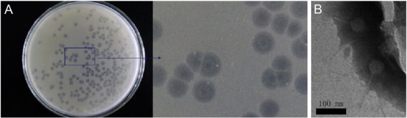
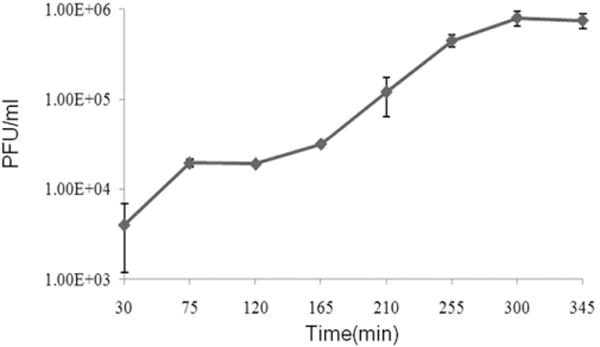
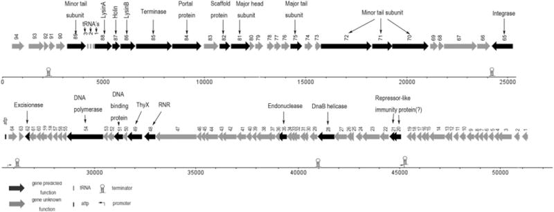
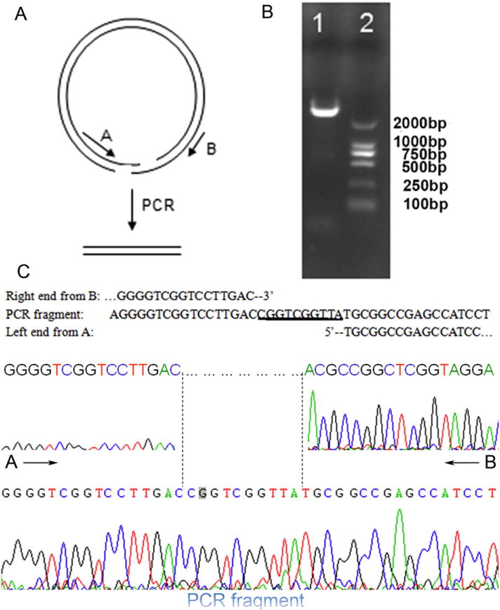
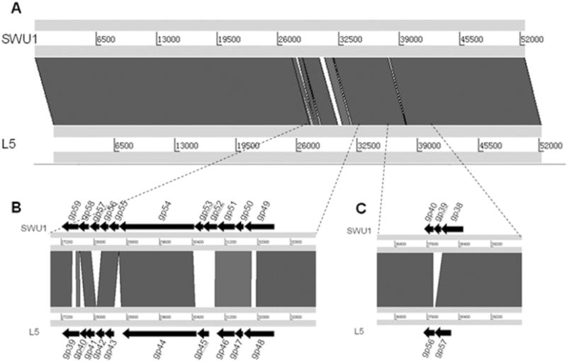
Mycobacteriophage SWU1 is a newly isolated phage from soil sample collected in Sichuan province, China using Mycobacterium smegmatis mc(2)155 as host. Plaque, phage morphology and one-step growth curve were characterized. The complete genomic sequence of phage SWU1 was determined by shotgun sequencing. The ends of SWU1 were determined. Structural proteins of SWU1 were analyzed by NanoLC-ESI-MS/MS. Seven ORFs were identified as structural protein encoded by SWU1 genome. The genetic basis underlying the SWU1 plaque was explored using comparative genomics. Prophages homologous to SWU1 were identified in two pathogens, Segniliparus rugosus ATCC BAA-974 and Mycobacterium rhodesiae JS60. Genus Segniliparus is a member of the order Corynebacteriales. To our knowledge, this is the first report of Mycobacterium prophages in different genera.
Keywords: Electron microscopy; Genome; Mycobacteriophage; Plaque; Proteome.
Copyright © 2015. Published by Elsevier B.V.
Figures
Similar articles
-
Biology of a novel mycobacteriophage, SWU1, isolated from Chinese soil as revealed by genomic characteristics.J Virol. 2012 Sep;86(18):10230-1. doi: 10.1128/JVI.01568-12. J Virol. 2012. PMID: 22923793 Free PMC article.
-
Genomic and proteomic portrait of a novel mycobacteriophage SWU2 isolated from China.Infect Genet Evol. 2021 Jan;87:104665. doi: 10.1016/j.meegid.2020.104665. Epub 2020 Dec 3. Infect Genet Evol. 2021. PMID: 33279716
-
Complete genome sequence analysis of the novel mycobacteriophage Shandong1.Arch Virol. 2017 Dec;162(12):3903-3905. doi: 10.1007/s00705-017-3534-7. Epub 2017 Aug 21. Arch Virol. 2017. PMID: 28828700
-
On the nature of mycobacteriophage diversity and host preference.Virology. 2012 Dec 20;434(2):187-201. doi: 10.1016/j.virol.2012.09.026. Epub 2012 Oct 22. Virology. 2012. PMID: 23084079 Free PMC article. Review.
-
Mycobacteriophages: genes and genomes.Annu Rev Microbiol. 2010;64:331-56. doi: 10.1146/annurev.micro.112408.134233. Annu Rev Microbiol. 2010. PMID: 20528690 Review.
Cited by
-
Complete genome sequence analysis of a new Escherichia phage, GaoY1-9D.Arch Virol. 2025 Apr 30;170(6):117. doi: 10.1007/s00705-025-06298-2. Arch Virol. 2025. PMID: 40304807
-
A novel temperate phage, vB_PstS-pAN, induced from the naphthalene-degrading bacterium Pseudomonas stutzeri AN10.Arch Virol. 2021 Aug;166(8):2267-2272. doi: 10.1007/s00705-021-05098-8. Epub 2021 May 18. Arch Virol. 2021. PMID: 34008105
-
Mycobacterium tuberculosis Rv2617c is involved in stress response and phage infection resistance.Heliyon. 2024 Mar 5;10(5):e27400. doi: 10.1016/j.heliyon.2024.e27400. eCollection 2024 Mar 15. Heliyon. 2024. PMID: 38495141 Free PMC article.
-
Mycobacteriophages and Their Applications.Antibiotics (Basel). 2024 Sep 27;13(10):926. doi: 10.3390/antibiotics13100926. Antibiotics (Basel). 2024. PMID: 39452193 Free PMC article. Review.
-
Isolation and characterization of a novel mycobacteriophage Kashi-VT1 infecting Mycobacterium species.Front Cell Infect Microbiol. 2023 Jul 21;13:1173894. doi: 10.3389/fcimb.2023.1173894. eCollection 2023. Front Cell Infect Microbiol. 2023. PMID: 37545854 Free PMC article.
References
-
- Bayer ME, Thurow H, Bayer MH. Penetration of the polysaccharide capsule of Escherichia coli (Bi161/42) by bacteriophage K29. Virology. 1979;94:95–118. - PubMed
-
- Butler W, Floyd MM, Brown JM, Toney SR, Daneshvar MI, Cooksey RC, Carr J, Steigerwalt AG, Charles N. Novel mycolic acid-containing bacteria in the family Segniliparaceae fam.nov., including the genus Segniliparus gen. nov., with descriptions of Segniliparus rotundus sp.nov and Segniliparus rugosus sp.nov. Int J Syst Evol Microbiol. 2005;55:1615–1624. - PubMed
-
- Curry E, Yehia M, Roberts S. CAPD peritonitis caused by Mycobacterium rhodesiae. Perit Dial Int. 2008;28:97–99. - PubMed
Publication types
MeSH terms
Substances
Grants and funding
LinkOut - more resources
Full Text Sources
Other Literature Sources
Molecular Biology Databases
