. 2015 May 15;24(10):2873-83.
doi: 10.1093/hmg/ddv049. Epub 2015 Feb 20.

The E3 ubiquitin ligase TRIM32 regulates myoblast proliferation by controlling turnover of NDRG2

Affiliations

The E3 ubiquitin ligase TRIM32 regulates myoblast proliferation by controlling turnover of NDRG2

Ekaterina I Mokhonova et al. Hum Mol Genet. .

Abstract

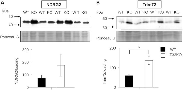
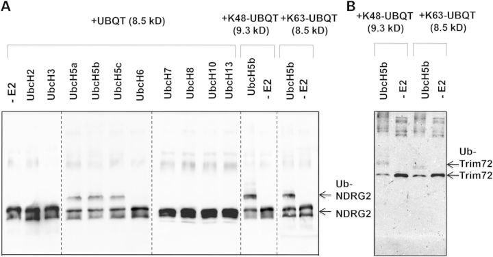
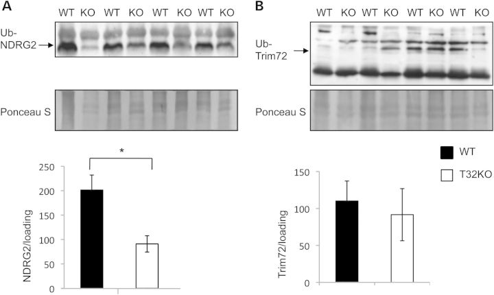
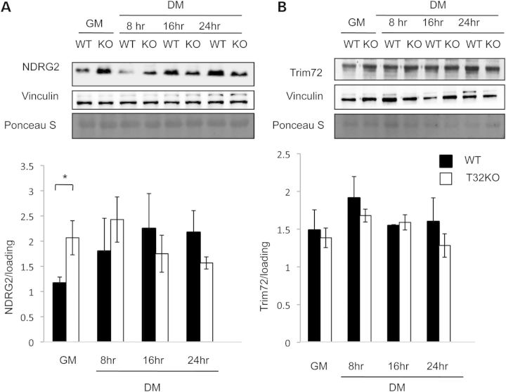
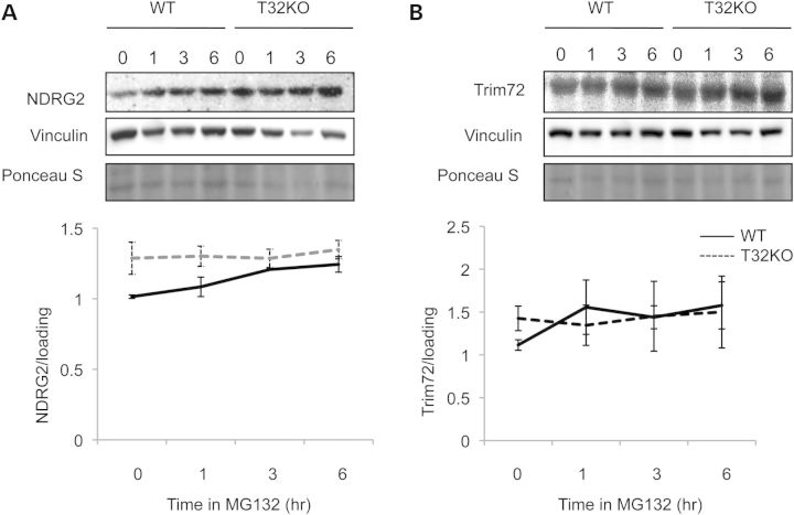
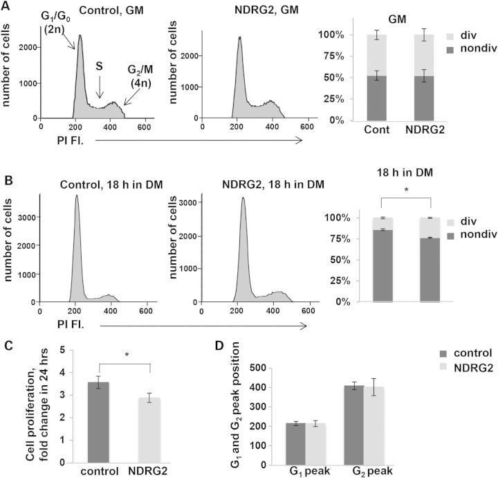
Limb girdle muscular dystrophy 2H is caused by mutations in the gene encoding the E3 ubiquitin ligase, TRIM32. Previously, we generated and characterized a Trim32 knockout mouse (T32KO) that displays both neurogenic and myopathic features. The myopathy in these mice is attributable to impaired muscle growth, associated with satellite cell senescence and premature sarcopenia. This satellite cell senescence is due to accumulation of the SUMO ligase PIASy, a substrate of TRIM32. The goal of this investigation was to identify additional substrates of TRIM32 using 2D fluorescence difference gel electrophoresis (2D-DIGE) in order to further explore its role in skeletal muscle. Because TRIM32 is an E3 ubiquitin ligase, we reasoned that TRIM32's substrates would accumulate in its absence. 2D-DIGE identified 19 proteins that accumulate in muscles from the T32KO mouse. We focused on two of these proteins, NDRG2 and TRIM72, due to their putative roles in myoblast proliferation and myogenesis. Follow-up analysis confirmed that both proteins were ubiquitinated by TRIM32 in vitro; however, only NDRG2 accumulated in skeletal muscle and myoblasts in the absence of TRIM32. NDRG2 overexpression in myoblasts led to reduced cell proliferation and delayed cell cycle withdrawal during differentiation. Thus, we identified NDRG2 as a novel target for TRIM32; these findings further corroborate the hypothesis that TRIM32 is involved in control of myogenic cells proliferation and differentiation.


Figures

Figure 1.
Figure 1.
Levels of NDRG2 and Trim72 proteins are elevated in total muscle lysates of WT and T32KO mice. Western blot analysis of NDRG2 and Trim72 proteins expressed in gastrocnemius from WT and T32KO muscles. Gastrocnemius western blot was stained for NDRG2 with NDRG2 Ab (A) or for Trim72 with Trim72 Ab (B); 40 mkg of total protein was loaded per lane. Samples from four animals per group were analyzed and representative blots are shown. The Ponceau S stained blots are shown as a loading control. Densitometry data normalized to loading, data are shown as mean ± SEM (*P < 0.05). Statistical analysis by t-test.
Figure 2.
Figure 2.
Trim32 is able to ubiquitinate recombinant NDRG2 and Trim72 proteins in vitro. (A) Trim32 ubiquitinated NDRG2 in conjunction with UbcH5a, UbcH5b and UbcH5C. Recombinant Trim32 was incubated in the presence of ATP, ubiquitin, E1 and several different E2 s as specified in the figure. Lane (–E2) indicates no E2 was added. The panel shows blot stained with anti-NDRG2 Ab. (B) Trim32 ubiquitinated Trim72 in conjunction with UbcH5b E2. The panel shows blot stained with anti-Trim72 Ab. Trim32 specifically ubiquitinated NDRG2 and TRim72 as evidenced by ∼10 kDa increment shifts in the molecular mass of the target proteins.
Figure 3.
Figure 3.
Levels of NDRG2 protein are significantly reduced in ubiquitinated fractions from T32KO muscles. (A and B) Western blot analysis of levels of NDRG2 and Trim 72 proteins in ubiquitinated fractions eluted from ubiquitin-affinity resin. Total muscle lysates from T32KO and WT muscles were incubated with ubiquitin-affinity resin (anti-ubiquitin monoclonal antibody (clone FK2) covalently coupled to agarose gel, ‘MBL’) for 2 h at RT, extensively washed, and bound proteins were eluted by reducing sample buffer. Panel A shows blot stained with anti-NDRG2 Ab, and panel B shows blot stained with anti-Trim72 Ab. Samples from four animals per group were analyzed and representative blots are shown. The Ponceau S stained blots are shown as a loading control. Densitometry data normalized to loading, data are shown as mean ± SEM (*P < 0.05). Statistical analysis by t-test.
Figure 4.
Figure 4.
NDRG2 levels increased in primary T32KO myoblasts comparing with WT cells. Western blot analysis demonstrates expression of NDRG2 (A) and Trim72 (B) proteins in primary myoblasts. Representative blots of total cell lysates are shown for each protein, in myogenic cells in GM and at different indicated times after switching to DM. Vinculin and Ponceau S stained blots are included to show the relative concentration of proteins in the cell lysates. Densitometry data were normalized to loading. Values represent the mean of at least three independent experiments ± SEM (*P < 0.05). Statistical analysis by t-test.
Figure 5.
Figure 5.
NDRG2 accumulates in WT myoblasts in the presence of proteasome inhibitor MG132. (A and B) Primary myogenic cells were treated with 50 μm of the inhibitor of proteasome activity, MG132, for 1, 3 or 6 h. Cells represented by the lane labeled ‘0’ were treated with dimethyl sulfoxide (vehicle) alone for 6 h. NDRG2 accumulated in WT myoblasts in the presence of MG132; in the T32KO cells the levels of NDRG2 were maintained, independent of the presence of MG13. Anti-vinculin blots and Ponceau S stained blots are shown as a loading control. Densitometry data normalized to loading and shown as mean ± SEM. Statistical analysis by t-test.
Figure 6.
Figure 6.
Over-expression of NDRG2 causes a delay in cell cycle withdrawal and slows down cell proliferation. (A and B) Cell cycle analysis of C2C12 myoblasts over-expressing NDRG2. C2C12 myoblasts were stained with PI and analyzed by flow cytometry. (A, B) Representative flow cytometric histograms showing the cell cycle distribution. (A) C2C12 control- and NDRG2-transduced cells in GM. (B) C2C12 control- and NDRG2-transduced cells were cultured for 18 h in DM. G1/G0, S and G2/M peaks are shown. Percentages of dividing (div) (S+G2/M) and non-dividing (nondiv.) (G1/G0) cells were calculated. Values represent the mean of at least three independent experiments ± SEM. (C) Effect of elevated NDRG2 levels on C2C12 myoblast proliferation. Cell proliferation index is presented as a fold change of the number of cells in 24 h. Data are summarized and presented as the mean of three independent experiments ± SEM (*P < 0.05). Statistical analysis by t-test. (D) Positions of G1 and G2 peaks are not changed on the PI fluorescence axis in NDRG2-transduced cells. Data are summarized and presented as the mean of three independent experiments ± SEM.

References

    1. Nigro V., Savarese M. (2014) Genetic basis of limb-girdle muscular dystrophies: the 2014 update. Acta Myol., 33, 1–12. - PMC - PubMed
    1. Frosk P., Weiler T., Nylen E., Sudha T., Greenberg C.R., Morgan K., Fujiwara T.M., Wrogemann K. (2002) Limb-girdle muscular dystrophy type 2H associated with mutation in TRIM32, a putativeE3-ubiquitin-ligase gene. Am. J. Hum. Genet., 70, 663–672. - PMC - PubMed
    1. Reymond A., Meroni G., Fantozzi A., Merla G., Cairo S., Luzi L., Riganelli D., Zanaria E., Messali S., Cainarca S., et al. (2001) The tripartite motif family Identifies cell compartments. EMBO J., 20, 2140–2151. - PMC - PubMed
    1. Saccone V., Palmieri M., Passamano L., Piluso G., Meroni G., Politano L., Nigro V. (2008) Mutations that impair interaction properties of TRIM32 associated with limb-girdle muscular dystrophy 2H. Hum. Mutat., 29, 240–247. - PubMed
    1. Cossee M., Lagier-Tourenne C., Seguela C., Mohr M., Leturcq F., Gundesli H., Chelly J., Tranchant C., Koenig M., Mandel J.L. (2009) Use of SNP array analysis to identify a novel TRIM32 mutation in limb-girdle muscular dystrophy type 2H. Neuromuscul. Disord., 19, 255–260. - PubMed

Publication types