

Autonomic dysfunction syndromes after acute brain injury
- PMID: 25701906
- PMCID: PMC8101086
- DOI: 10.1016/B978-0-444-63521-1.00034-0
Autonomic dysfunction syndromes after acute brain injury
Abstract
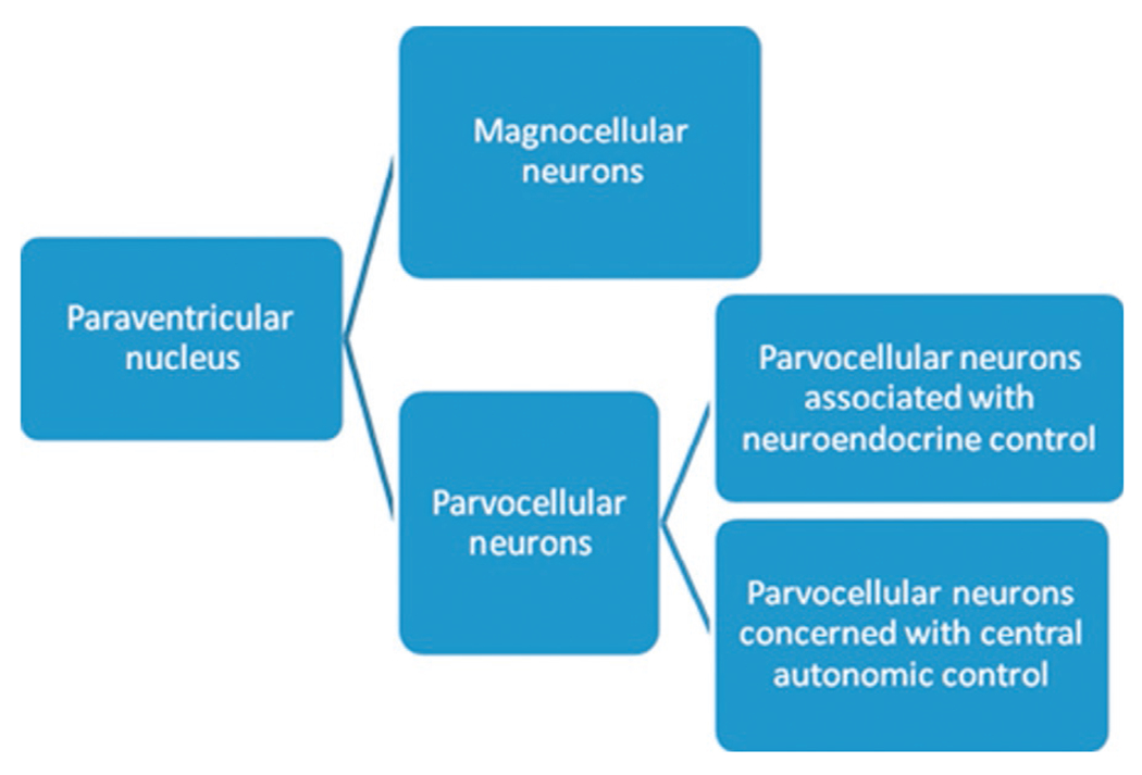
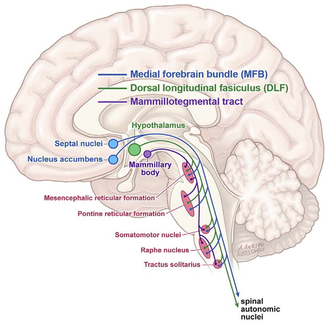
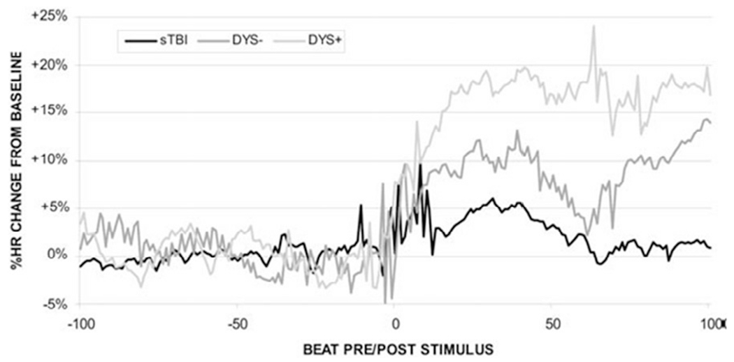
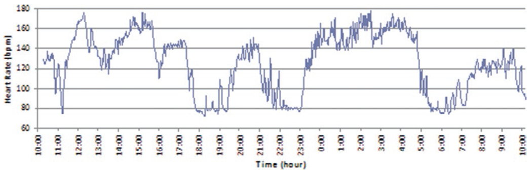
The central autonomic nervous system (CAN) is a multifaceted, richly connected neural network incorporating the hypothalamus, its descending tracts through the brainstem, the insular cortex and down into the spinal cord. All levels of the CAN are susceptible to injury following traumatic brain injury (TBI), whether from focal or diffuse injury. Focal injuries would be expected to produce localized damage to CAN control centers, whereas the effects of diffuse injuries are presumed to be more diverse and/or widely distributed. As the combination of focal and diffuse injury following TBI can vary widely from one individual to the next, the impact of focal injuries is best understood with reference to the focal ischemic stroke literature. Subarachnoid hemorrhage (SAH), a common complication following TBI, also has predictable effects on autonomic control that can be understood with reference to spontaneous SAH literature. Finally, paroxysmal sympathetic hyperactivity (PSH), a syndrome incorporating episodes of heightened sympathetic drive and motor overactivity following minor stimulation, is discussed as an example of what happens when central inhibitory control of spinal cord autonomics is impaired.
Keywords: central autonomic network; hyperadrenergic crisis; paroxysmal sympathetic hyperactivity; stroke; subarachnoid hemorrhage; traumatic brain injury.
© 2015 Elsevier B.V. All rights reserved.
Figures
Similar articles
-
Autonomic dysfunction following traumatic brain injury: translational insights.Neurosurg Focus. 2019 Nov 1;47(5):E8. doi: 10.3171/2019.8.FOCUS19517. Neurosurg Focus. 2019. PMID: 31675718 Review.
-
Paroxysmal sympathetic hyperactivity in pediatric traumatic brain injury: A case series of four patients.Auton Neurosci. 2015 Dec;193:149-51. doi: 10.1016/j.autneu.2015.08.003. Epub 2015 Aug 6. Auton Neurosci. 2015. PMID: 26277041
-
Paroxysmal sympathetic hyperactivity during traumatic brain injury.Clin Neurol Neurosurg. 2022 Jan;212:107081. doi: 10.1016/j.clineuro.2021.107081. Epub 2021 Nov 27. Clin Neurol Neurosurg. 2022. PMID: 34861468 Review.
-
Paroxysmal sympathetic hyperactivity: the storm after acute brain injury.Lancet Neurol. 2017 Sep;16(9):721-729. doi: 10.1016/S1474-4422(17)30259-4. Lancet Neurol. 2017. PMID: 28816118 Review.
-
Paroxysmal Sympathetic Hyperactivity.Semin Neurol. 2020 Oct;40(5):485-491. doi: 10.1055/s-0040-1713845. Epub 2020 Sep 9. Semin Neurol. 2020. PMID: 32906174 Review.
Cited by
-
Placebo Effects in Traumatic Brain Injury.J Neurotrauma. 2018 Jun 1;35(11):1205-1212. doi: 10.1089/neu.2017.5506. Epub 2018 Apr 5. J Neurotrauma. 2018. PMID: 29343158 Free PMC article. Review.
-
Age-related changes in baroreflex sensitivity and cardiac autonomic tone in children mirrored by regional brain gray matter volume trajectories.Pediatr Res. 2018 Feb;83(2):498-505. doi: 10.1038/pr.2017.273. Epub 2017 Dec 20. Pediatr Res. 2018. PMID: 29261644 Free PMC article.
-
Quantitative susceptibility mapping at 7 T in COVID-19: brainstem effects and outcome associations.Brain. 2024 Dec 3;147(12):4121-4130. doi: 10.1093/brain/awae215. Brain. 2024. PMID: 39375207 Free PMC article.
-
Hypotension and Adverse Outcomes in Moderate to Severe Traumatic Brain Injury: A Systematic Review and Meta-Analysis.JAMA Netw Open. 2024 Nov 4;7(11):e2444465. doi: 10.1001/jamanetworkopen.2024.44465. JAMA Netw Open. 2024. PMID: 39527054 Free PMC article.
-
Clinical Potential of Immunotherapies in Subarachnoid Hemorrhage Treatment: Mechanistic Dissection of Innate and Adaptive Immune Responses.Aging Dis. 2023 Oct 1;14(5):1533-1554. doi: 10.14336/AD.2023.0126. Aging Dis. 2023. PMID: 37196120 Free PMC article. Review.
References
-
- Anderson VL, Ahmed G, Duraski SA et al. (2004). Alternative treatment in the management of combined hyperadrenergia and spasticity in the adult with a severe traumatic brain injury: case report. Arch Phys Med Rahabil 85: e15.
-
- Badjatia N (2009). Hyperthermia and fever control in brain injury. Crit Care Med 37: S250–S257. - PubMed
-
- Badjatia N, Fernandez L, Schmidt JM et al. (2010). Impact of induced normothermia on outcome after subarachnoid hemorrhage. Neurosurg 66: 696–701. - PubMed
-
- Baguley IJ (2008a). The excitatory:inhibitory model (EIR model): an integrative explanation of acute autonomic overactivity syndromes. Med Hypotheses 70: 26–35. - PubMed
-
- Baguley IJ (2008b). Autonomic complications following central nervous system injury. Sem Neurol 28: 716–725. - PubMed
Publication types
MeSH terms
Grants and funding
LinkOut - more resources
Full Text Sources
Medical
Research Materials
