Review
. 2015:127:45-66.
doi: 10.1016/B978-0-444-52892-6.00004-0.

The neuropathology of traumatic brain injury

Affiliations
Review

The neuropathology of traumatic brain injury

Ann C Mckee et al. Handb Clin Neurol. 2015.

Abstract

Traumatic brain injury, a leading cause of mortality and morbidity, is divided into three grades of severity: mild, moderate, and severe, based on the Glasgow Coma Scale, the loss of consciousness, and the development of post-traumatic amnesia. Although mild traumatic brain injury, including concussion and subconcussion, is by far the most common, it is also the most difficult to diagnose and the least well understood. Proper recognition, management, and treatment of acute concussion and mild traumatic brain injury are the fundamentals of an emerging clinical discipline. It is also becoming increasingly clear that some mild traumatic brain injuries have persistent, and sometimes progressive, long-term debilitating effects. Evidence indicates that a single traumatic brain injury can precipitate or accelerate multiple age-related neurodegenerations, increase the risk of developing Alzheimer's disease, Parkinson's disease, and motor neuron disease, and that repetitive mild traumatic brain injuries can provoke the development of a tauopathy, chronic traumatic encephalopathy. Clinically, chronic traumatic encephalopathy is associated with behavioral changes, executive dysfunction, memory loss, and cognitive impairments that begin insidiously and progress slowly over decades. Pathologically, chronic traumatic encephalopathy produces atrophy of the frontal and temporal lobes, thalamus, and hypothalamus, septal abnormalities, and abnormal deposits of hyperphosphorylated tau (τ) as neurofibrillary tangles and disordered neurites throughout the brain. The incidence and prevalence of chronic traumatic encephalopathy and the genetic risk factors critical to its development are currently unknown. Chronic traumatic encephalopathy frequently occurs as a sole diagnosis, but may be associated with other neurodegenerative disorders, including Alzheimer's disease, Lewy body disease, and motor neuron disease. Currently, chronic traumatic encephalopathy can be diagnosed only at autopsy; however, promising efforts to develop imaging, spinal fluid, and peripheral blood biomarkers are underway to diagnose and monitor the course of disease in living subjects.

Keywords: Axonal injury; blast injury; brain trauma; concussion; mild traumatic brain injury; motor neuron disease; post-traumatic neurodegeneration; tau protein.


Figures

Fig. 4.1
Fig. 4.1
Neuropathologic changes associated with blast injury. (A–C) Subcortical frontal white matter shows loss of myelinated fibers, abnormal myelin clumps (asterisks), astrocytosis (arrows), luxol fast blue-hematoxylin and eosin stain, original magnification (A)×200, (B)×400, (C)×600. (D) Axons in the frontal white matter are severely depleted with many irregular axonal swellings, SMI-34 immunostaining, original magnification×400. (E) APP immunostaining shows axon retraction bulbs and other axonal irregularities, APP immunostain, original magnification×400. (F) Axonal swellings, SMI-34 immunostain, original magnification×600. (G) A single focus of perivascular neurofibrillary tangles (NFTs) and neurites at the depth of sulcus in inferior parietal cortex consistent with stage I/IV chronic traumatic encephalopathy (CTE), AT8 immunostaining for hyperphosphorylated tau, original magnification×100. (H) Perivascular NFTs at the sulcal depths of frontal cortex, AT8 immunostaining, original magnification×200. (I) Multiple areas of perivascular NFTs, AT8 immunostaining, original magnification×100. (J) Small blood vessels in the thalamus show prominent perivascular lymphocytic cuffing (asterisk) while a neighboring medium size artery shows focal degenerative calcification of the blood vessel wall (arrow), luxol fast blue-hematoxylin and eosin stain, original magnification×200. (K) A medium size vessel in the thalamus shows lymphocytic infiltration of the vascular wall and hemosiderin-laden macrophages (asterisk), luxol fast blue-hematoxylin and eosin stain, original magnification×400. (L) Focus of dystrophic calcification in the white matter, luxol fast blue-hematoxylin and eosin stain, original magnification×400. (Adapted from McKee and Robinson, 2014.)
Fig. 4.2
Fig. 4.2
Gross neuropathologic features of chronic traumatic encephalopathy (CTE). In advanced CTE, there is often diffuse cerebral atrophy, marked atrophy of the medial temporal lobe structures (hippocampus, amygdala and entorhinal cortex), enlargement of the lateral and third ventricles (with disproportionate enlargement of the third ventricle), and atrophy of the diencephalon and mammillary bodies. Other features include cavum septum pellucidum ((A) and (B)), fenestrated or torn posterior septum pellucidum (C–F, I), pallor of the substantia nigra (G, H) and pallor of the locus coeruleus (H).
Fig. 4.3
Fig. 4.3
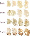
Staging of hyperphosphorylated tau pathology in chronic traumatic encephalopathy (CTE). In stage I CTE, hyperphosphorylated p-tau pathology is restricted to discrete foci in the cerebral cortex, most commonly in the superior, dorsolateral or lateral frontal cortices, and typically around small vessels at the depths of sulci. In stage II CTE, there are multiple epicenters at the depths of the cerebral sulci and spread of neurofibrillary pathology to the superficial layers of adjacent cortex. The medial temporal lobe is spared neurofibrillary p-tau pathology in stage II CTE although it becomes progressively more involved as disease severity increases. In stage III, p-tau pathology is widespread; the frontal, insular, temporal, and parietal cortices, amygdala, hippocampus and entorhinal cortex show widespread neurofibrillary pathology. In stage IV CTE, there is widespread severe p-tau pathology affecting most regions of the cerebral cortex and the medial temporal lobe, sparing calcarine cortex in all but the most severe cases. All images, CP-13 immunostained 50 μ tissue sections. Adapted from McKee et al., 2013.)
Fig. 4.4
Fig. 4.4
Microscopic features of stage IV chronic traumatic encephalopathy (CTE). (Top row) Whole mount coronal sections of the brain from cognitively intact 65 year old subject; CP-13 immunostained 50 μ tissue sections. (Second row) Whole mount 50 μ sections of brain from 66-year-old with stage IV CTE. There is widespread p-tau immunoreactive neurofibrillary pathology. (Third row) Microscopic sections from 66-year-old with stage IV CTE show irregular focal patches of dense p-tau pathology, centered around small blood vessels and most severe at the depths of the sulci; CP-13 immunostaining, 10 μm sections. (Fourth row) P-TDP-43 immunoreactive neuronal and glial inclusions and neurites are densely deposited in the lower layers of the frontal and temporal cortices of the 66-year-old with stage IV CTE; pTDP-43 immunostaining, 10 μm sections. There is also a slight tendency for the abnormal pTDP-43 deposits to be concentrated around small blood vessels.
Fig. 4.5
Fig. 4.5
Chronic traumatic encephalopathy (CTE) stage II–III and motor neuron disease. (Top row) Whole mount 50 μ sections of brain from 43-year-old with stage II–III CTE with motor neuron disease. P-tau immunoreactive neurofibrillary pathology is most dense in multiple foci at the depths of the sulci in the frontal lobes. There is also p-tau neurofibrillary pathology in the hippocampus and entorhinal cortex. (Second row) Microscopic sections show irregular focal patches of dense p-tau pathology in the frontal cortex, centered around small blood vessels and most severe at the depths of the sulci, as well as dense NFTs in CA1 of the hippocampus; CP-13 immunostaining, 50 μm free-floating sections. (Third row) P-TDP-43 immunoreactive neuronal and glial inclusions and neurites are densely deposited around blood vessels and in the subpial regions at the depths of the sulci of the frontal and temporal cortices; dense pTDP43 neuronal and glial inclusions and neurites are found in the spinal cord as well as the cerebral cortex; pTDP-43 immunostaining, 10 μm sections.

Similar articles

Cited by

References

    1. Aoki Y, Inokuchi R, Gunshin M, et al. Diffusion tensor imaging studies of mild traumatic brain injury: a meta-analysis. J Neurol Neurosurg Psychiatry. 2012;83:870–876. - PMC - PubMed
    1. Baker M, Litvan I, Houlden H, et al. Association of an extended haplotype in the tau gene with progressive supranuclear palsy. Hum Mol Genet. 1999;8:711–715. - PubMed
    1. Baugh CM, Stamm JM, Riley DO, et al. Chronic traumatic encephalopathy: neurodegeneration following repetitive concussive and subconcussive brain trauma. Brain Imaging Behav. 2012;6:244–254. - PubMed
    1. Bekris LM, Mata IF, Zabetian CP. The genetics of Parkinson disease. J Geriatr Psychiatry Neurol. 2010;23:228–242. - PMC - PubMed
    1. Belli S, Vanacore N. Proportionate mortality of Italian soccer players: is amyotrophic lateral sclerosis an occupational disease? Eur J Epidemiol. 2005;20:237–242. - PubMed

Publication types