. 2015 May;27(5):997-1007.
doi: 10.1016/j.cellsig.2015.02.012. Epub 2015 Feb 20.

Activation of β2-adrenergic receptor by (R,R')-4'-methoxy-1-naphthylfenoterol inhibits proliferation and motility of melanoma cells

Affiliations

Activation of β2-adrenergic receptor by (R,R')-4'-methoxy-1-naphthylfenoterol inhibits proliferation and motility of melanoma cells

Artur Wnorowski et al. Cell Signal. 2015 May.

Abstract

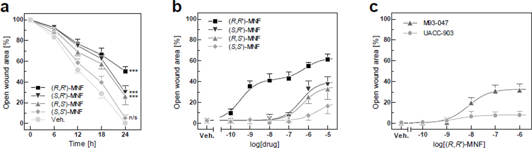
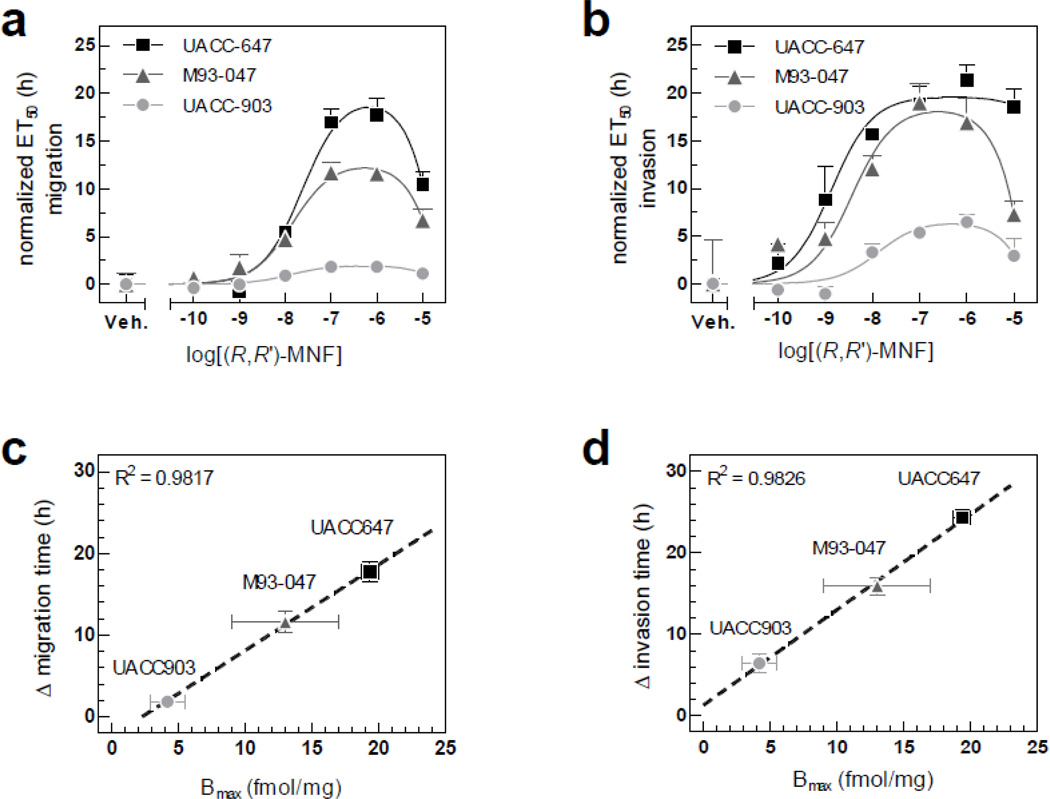
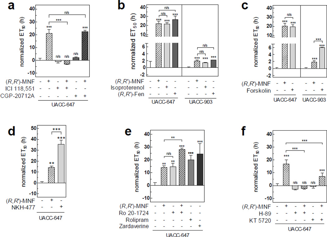
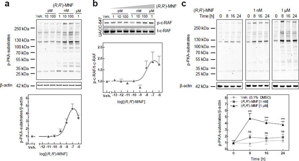
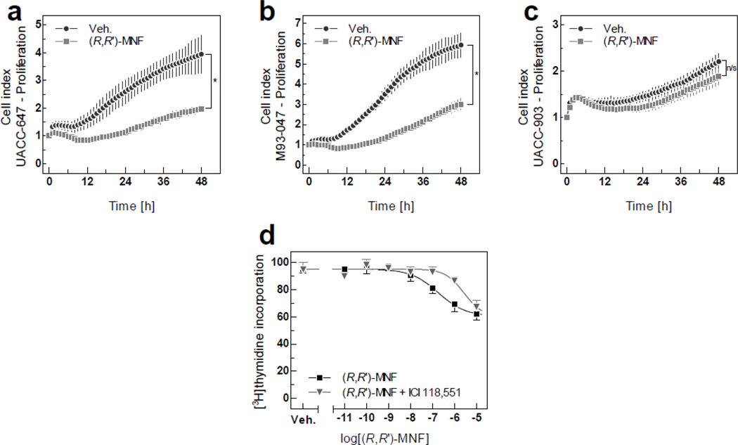
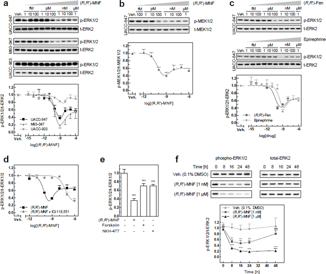
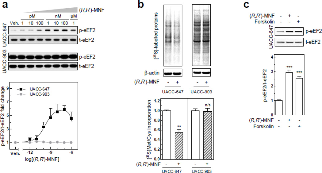
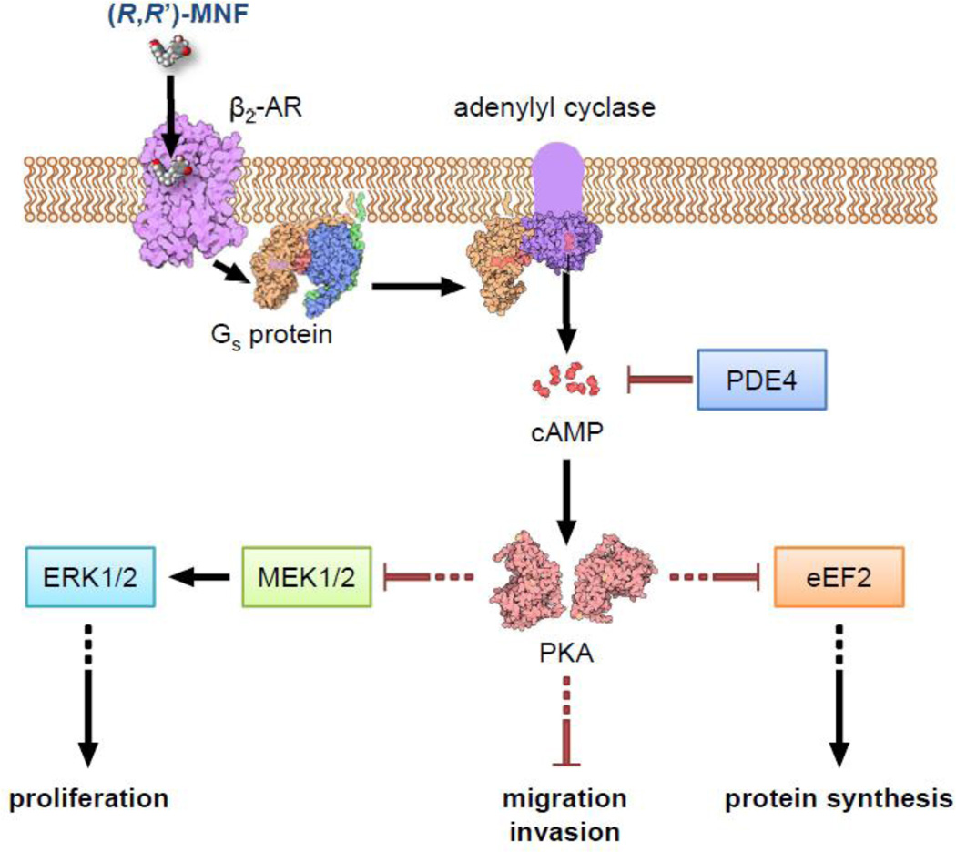
(R,R')-4'-methoxy-1-naphthylfenoterol [(R,R')-MNF] is a highly-selective β2 adrenergic receptor (β2-AR) agonist. Incubation of a panel of human-derived melanoma cell lines with (R,R')-MNF resulted in a dose- and time-dependent inhibition of motility as assessed by in vitro wound healing and xCELLigence migration and invasion assays. Activity of (R,R')-MNF positively correlated with the β2-AR expression levels across tested cell lines. The anti-motility activity of (R,R')-MNF was inhibited by the β2-AR antagonist ICI-118,551 and the protein kinase A inhibitor H-89. The adenylyl cyclase activator forskolin and the phosphodiesterase 4 inhibitor Ro 20-1724 mimicked the ability of (R,R')-MNF to inhibit migration of melanoma cell lines in culture, highlighting the importance of cAMP for this phenomenon. (R,R')-MNF caused significant inhibition of cell growth in β2-AR-expressing cells as monitored by radiolabeled thymidine incorporation and xCELLigence system. The MEK/ERK cascade functions in cellular proliferation, and constitutive phosphorylation of MEK and ERK at their active sites was significantly reduced upon β2-AR activation with (R,R')-MNF. Protein synthesis was inhibited concomitantly both with increased eEF2 phosphorylation and lower expression of tumor cell regulators, EGF receptors, cyclin A and MMP-9. Taken together, these results identified β2-AR as a novel potential target for melanoma management, and (R,R')-MNF as an efficient trigger of anti-tumorigenic cAMP/PKA-dependent signaling in β2-AR-expressing lesions.

Keywords: Beta blocker; Beta2 adrenoreceptor selective agonist; Melanocortin 1 receptor; Melanocyte; Metastasis; Migration.


Conflict of interest statement

Conflict of interest

The authors state no conflict of interest other than the fact that IW Wainer, M Bernier, RK Paul, LR Toll and LA Jimenez are listed as co-inventors on a patent application for the use of fenoterol and other fenoterol derivatives [including (R,R’)-MNF] in the treatment of glioblastomas and astrocytomas. Wainer, Bernier and Paul have assigned their rights in the patent to the US government, but will receive a percentage of any royalties that may be received by the government.

Figures

Figure 1
Figure 1. (R,R’)-MNF inhibits in vitro wound healing in cultured melanoma cells
(a) Scratch assays were performed with UACC-647 cells in the presence of (S,S’), (S,R’), (R,S’) and (R,R’) stereoisomers of MNF (1 µM) or vehicle (DMSO, 0.1%). Pictures were taken immediately after wound generation and every 6 h until the closure of the scratch wound in vehicle-treated cells (24 h). Open wound areas were measured over time and plotted. Values were gathered from three independent experiments carried out in quadruplicates (n = 12). Data are expressed as mean ± 95% confidence interval. Effects of MNF isomers versus control were statistically evaluated at 24 h time-point using one-way ANOVA and Tukey’s post-hoc test. ***, P < 0.001; n/s, not significant. (b) UACC-647 cells were subjected to scratch wound and treated with 100 pM, 1 nM, 10 nM, 100 nM, 1 µM and 10 µM of (R,R’), (S,R’), (R,S’) and (S,S’) stereoisomers of MNF or vehicle (DMSO, 0.1%). Dose-dependent effects of MNF stereoisomers were evaluated using non-linear regression (see section 3.1 for IC50 values). (c) Capacity of (R,R’)-MNF to inhibit wound healing was assessed in M93-047 and UACC-903 cells. The relative wound surface area of 12 independent observations at the 24 h time-point is plotted.
Figure 2
Figure 2. (R,R’)-MNF activity on melanoma cell migration depends on the expression of β2-AR
(a) Migration of UACC-647, M93-047 and UACC-903 melanoma cells was studied using xCELLigence system. Serum-depleted cells were treated with increasing concentrations of (R,R’)-MNF (from 100 pM to 10 µM) or vehicle (DMSO, 0.1%) and allowed to migrate via microporous PET membrane towards 10% FBS. CI was recorded over time (see Fig. S2) and was used to calculate ET50 values. All ET50 values were normalized to the data obtained for vehicle-treated cells. Bell-shaped curve was fitted to the data and IC50 values were calculated. (b) Invasion of UACC-647, M93-047 and UACC-903 cells through Matrigel coating was studied in parallel using the same approach. (c) Linear regression model was used to correlate maximal delay in migration time (Δ migration time) caused by (R,R’)-MNF with expression level of β2-AR (Bmax). (d) Analogously, (R,R’)-MNF-dependent delay in invasion through Matrigel was correlated with expression level of β2-AR.
Figure 3
Figure 3. (R,R’)-MNF acts via cAMP to inhibit melanoma cell migration
UACC-647 or UACC-903 melanoma cells were serum-starved for 20 h and their migratory capabilities were assessed using xCELLigence system. Recorded CI values that were used to calculate ET50 values are depicted on Fig. S5. (a) Serum-starved UACC-647 cells were pre-treated with ICI-118,551 (50 nM) or CGP-20712A (50 nM) for 45 min followed by the addition of (R,R’)-MNF (100 nM) or vehicle (DMSO, 0.1%) and allowed to migrate via PET membrane towards 10% FBS. Serum-free medium was used as negative control (Neg. ctrl.). ET50 was calculated for each treatment and normalized to vehicle. (b) Serum-depleted UACC-647 and UACC-903 cells were treated with β2-AR agonists: (R,R’)-MNF, isoproterenol or (R,R’)-Fen (all at 100 nM concentration). Migration through PET membrane was measured. (c) Activity of forskolin (10 µM) was compared with the anti-migratory properties of (R,R’)-MNF in UACC-647 and UACC-903 cells. Normalized ET50 values are given. (d) UACC-647 cells were serum-starved and treated with NKH-477 (10 µM), (R,R’)-MNF or vehicle (DMSO, 0.1%). (e) UACC-647 cells were incubated with (R,R’)-MNF (100 nM), Ro 20-1724 (10 µM) or combination of the two. Additionally, the cells were treated with Rolipram (10 µM) or Zardaverine (10 µM). ET50 values were calculated. (f) UACC-647 cells were serum-depleted and pre-treated with H-89 (20 µM) or KT 5720 (10 µM) followed by the addition of (R,R’)-MNF or vehicle (DMSO, 0.1%). Anti-migratory effect of tested compounds is presented as ET50 values. All experiments were performed in triplicates. Error bars represent SD. Statistical evaluation of either ‘treatments versus control cells’ (symbols directly above the bars) or ‘differences between the indicated treatments’ was performed using one-way ANOVA followed by Tukey’s post-hoc test. ***, P < 0.001; **, P < 0.01; n/s, not significant.
Figure 4
Figure 4. (R,R’)-MNF activates PKA
(a) UACC-647 cells were serum-starved for 3 h and treated for 15 min with increasing concentrations of (R,R’)-MNF (10 pM – 1 µM) or vehicle (DMSO, 0.1%). PKA activity was assessed by western blotting using antibody detecting phosphorylated substrates of PKA. β-actin was used as loading control. Intensities of all phospho-PKA substrate bands were measured, normalized to β-actin and plotted. (b) Serum-depleted UACC-647 cells were challenged with (R,R’)-MNF (1 pM - 1 µM) or vehicle (DMSO, 0.1%). Expression and phosphorylation pattern of c-Raf at inhibitory Ser259 was determined by western blotting. (c) Serum-starved UACC-647 cells were treated with (R,R’)-MNF (1 nM or 1 µM) or vehicle (DMSO, 0.1%) for 0, 8, 16 or 24 h. Levels of phosphorylation of PKA targets were plotted over time and statistically evaluated using two-way ANOVA and Bonferroni post-hoc test. ***, P < 0.001; n/s, not significant (versus control cells).
Figure 5
Figure 5. Anti-mitogenic activities of (R,R’)-MNF
Proliferation of human melanoma cell lines in culture was monitored in real-time using xCELLigence system. The UACC-647 (a), M93-047 (b) and UACC-903 (c) cells were cultured for 15 h followed by treatment with 100 nM (R,R’)-MNF or vehicle (DMSO, 0.1%). Impedance was recorded every 15 min., but to improve the clarity of the graphs only every fourth readout was plotted. Data show the average ± SD of three independent measurements. Differences between CI values for (R,R’)-MNF-treated and control cells were statistically evaluated using Student’s t-test. *, P < 0.05; n/s, not significant. (d) UACC-647 cells were pretreated with ICI-118,551 (100 nM) or vehicle (H2O) for 30 min followed by a 48-h incubation with increasing concentrations of (R,R’)-MNF. Cellular proliferation was measured after subsequent addition of [3H]thymidine for 4 h (see Materials and Methods for further details). Data show the average ± SD of three independent experiments, each performed in triplicate wells.
Figure 6
Figure 6. (R,R’)-MNF inhibits ERK activity
(a) UACC-647, M93-047 and UACC-903 cells were serum-starved for 3 h and subsequently treated with vehicle (DMSO, 0.1%) or a range of (R,R’)-MNF concentrations (1 fM to 1 µM) for 15 min. Cell lysates were immunoblotted for phosphorylated and total forms of ERK. Top panel depicts representative blots. Values on the graph (bottom panel) represent means ± SD from 3 independent experiments. (b) Serum-starved UACC-647 cells were treated with (R,R’)-MNF (100 fM to 1 µM) for 15 min; cell lysates were prepared and immunoblotted for total and phosphorylated forms of MEK1/2. Representative blots (top panel) and densitometric quantification (bottom panel) are given based on 3 independent experiments. (c) Serum-depleted UACC-647 cells were treated with vehicle (DMSO, 0.1%) or increasing concentrations of (R,R’)-Fen or epinephrine (1 fM to 1 µM) for 15 min. The levels of total and phosphorylated ERK were determined by immunoblotting (top panel) and dose-response curves were generated (bottom panel). (d) UACC-647 cells were serum-starved for 3 h followed by the addition of ICI-118,551 (50 nM) or vehicle (water, 0.1%) for 15 min. Subsequently, cells were treated with a range of (R,R’)-MNF concentrations (1 fM to 1 µM) or vehicle (DMSO, 0.1%). The levels of phosphorylated and total forms of ERK1/2 were determined by ELISA. (e) UACC-647 cells were serum-starved for 3 h and treated with (R,R’)-MNF (1 nM), forskolin (10 µM), NKH-477 (10 µM) or vehicle (DMSO, 0.1%) for 15 min. Expression of total and phosphorylated forms of ERK1/2 were assessed by ELISA. One-way ANOVA followed by Tukey’s post-hoc test were used to statistically evaluate the effect of the treatments versus control cells. ***, P < 0.001. (f) Serum-depleted UACC-647 cells were treated with (R,R’)-MNF (1 nM or 1 µM) or vehicle (DMSO, 0.1%) for 0, 8, 16, 24 or 48 h. Changes in ERK phosphorylation pattern were studied by western blotting. Representative blots are depicted. Densitometric quantitation of the blots was performed and plotted (bottom panel). Statistical analysis was performed applying two-way ANOVA and Bonferroni post-hoc test. Asterisk symbol depicts differences in phospho-ERK in (R,R’)-MNF-treated cells versus controls for each time-point. ***, P < 0.001; **, P < 0.01; n/s, not significant.
Figure 7
Figure 7
(a) UACC-647 and UACC-903 cells were serum-starved for 3 h and treated with vehicle (DMSO, 0.1%) or a range of (R,R’)-MNF concentrations (1 pM to 1 µM) for 15 min. Cell lysates were tested for the expression of phosphorylated and total forms of eEF2 by means of western blotting. Representative blots (top panel) and densitometric quantification (bottom panel) are depicted. (b) Serum-depleted UACC-647 and UACC-903 cells were treated with (R,R’)-MNF (100 nM) or vehicle (DMSO, 0.1%) in Met/Cys-free medium for 15 h followed by [35S] labeling for 30 min. De novo protein synthesis was assessed by autoradiography followed by the detection by immunoblotting. Effect of (R,R’)-MNF on the [35S]-Met/Cys incorporation was evaluated by Student’s t-test. **, P < 0.01; n/s, not significant. (c) UACC-647 cells were serum-starved for 3 h and then treated with vehicle (DMSO, 0.1%), (R,R’)-MNF (100 nM) or forskolin (10 µM) for 15 min. Cells were lysed and expression of phosphorylated and total forms of eEF2 was analyzed by western blotting. One-way ANOVA was used to evaluate the effect of the treatments versus control cells. ***, P < 0.001.
Figure 8
Figure 8. Mechanism of anti-tumorigenic activities of (R,R’)-MNF in melanoma cells
Binding of (R,R’)-MNF to β2-AR leads to cAMP accumulation via activation of adenylyl cyclase. The resultant activation of PKA promotes a cascade of downstream signaling pathways that leads to blunted proliferation, motility and protein synthesis in melanoma cells. Phosphodiesterases can reduce cAMP-dependent signaling. This figure and the graphical abstract are adapted from illustrations obtained from ‘Molecule of the Month’ by David S. Goodsell/RCSB PDB available under CC-BY-3.0 license.

Similar articles

Cited by

References

    1. Jozwiak K, Woo AY, Tanga MJ, Toll L, Jimenez L, Kozocas JA, Plazinska A, Xiao RP, Wainer IW. Bioorg Med Chem. 2010;18:728–736. - PMC - PubMed
    1. Toll L, Jimenez L, Waleh N, Jozwiak K, Woo AY, Xiao RP, Bernier M, Wainer IW. J Pharmacol Exp Ther. 2011;336:524–532. - PMC - PubMed
    1. Paul RK, Wnorowski A, Gonzalez-Mariscal I, Nayak SK, Pajak K, Moaddel R, Indig FE, Bernier M, Wainer IW. Biochem Pharmacol. 2014;87:547–561. - PMC - PubMed
    1. Yang G, Zhang G, Pittelkow MR, Ramoni M, Tsao H. J Invest Dermatol. 2006;126:2490–2506. - PubMed
    1. O’Leary AP, Fox JM, Pullar CE. J Cell Physiol. 2014 - PMC - PubMed

Publication types

MeSH terms