. 2015 Feb 26;160(5):829-841.
doi: 10.1016/j.cell.2015.01.033. Epub 2015 Feb 19.

Sensory detection of food rapidly modulates arcuate feeding circuits

Affiliations

Sensory detection of food rapidly modulates arcuate feeding circuits

Yiming Chen et al. Cell. .

Abstract

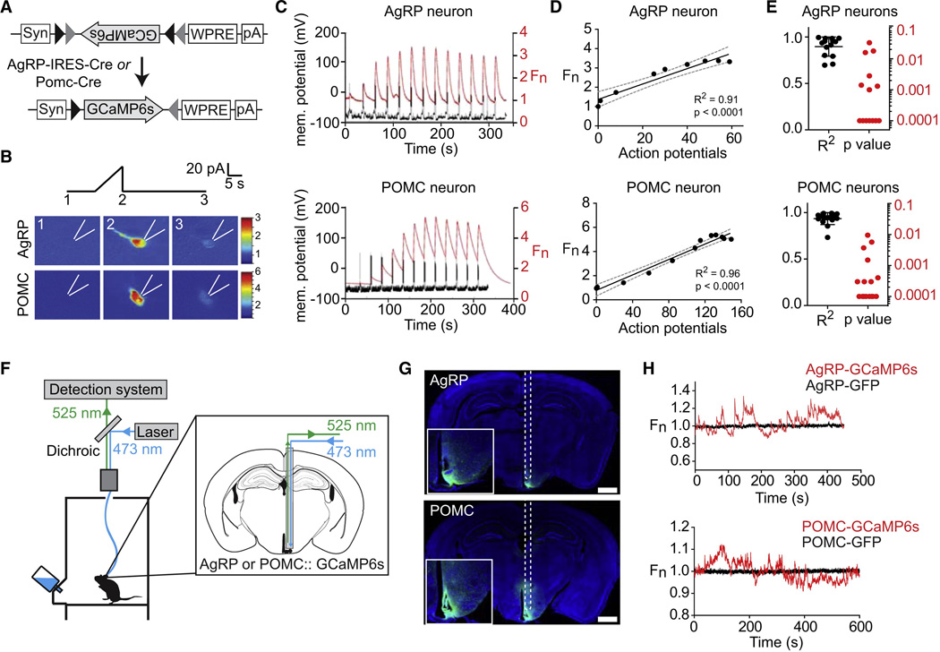
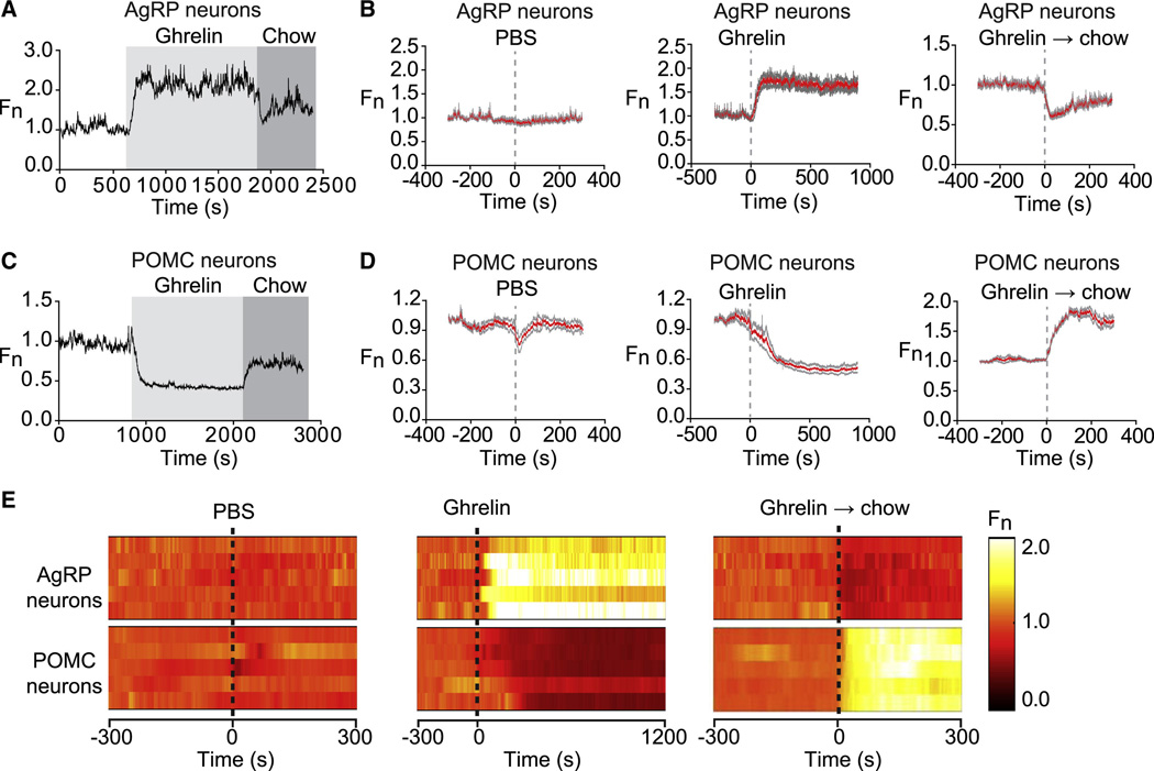
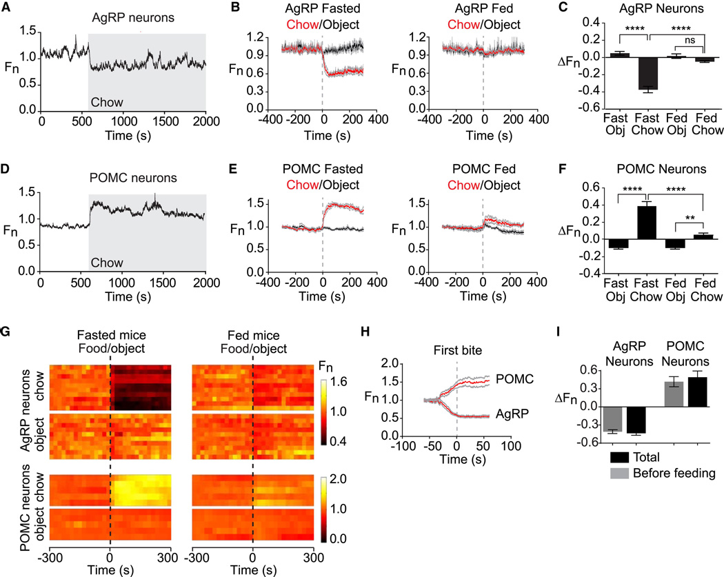
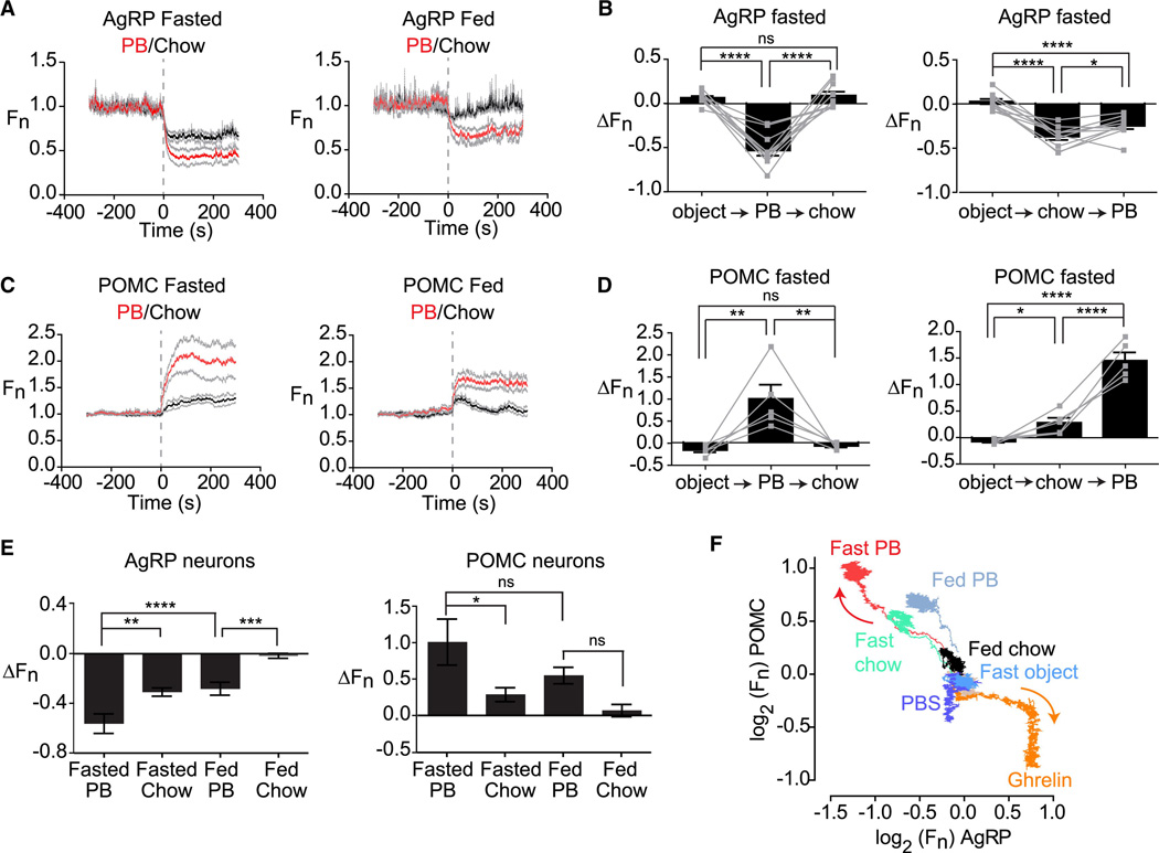
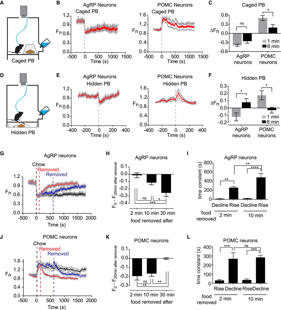
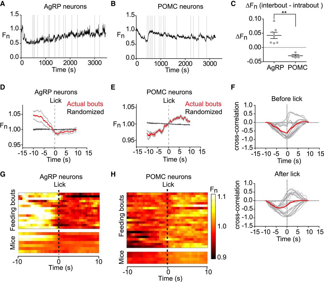
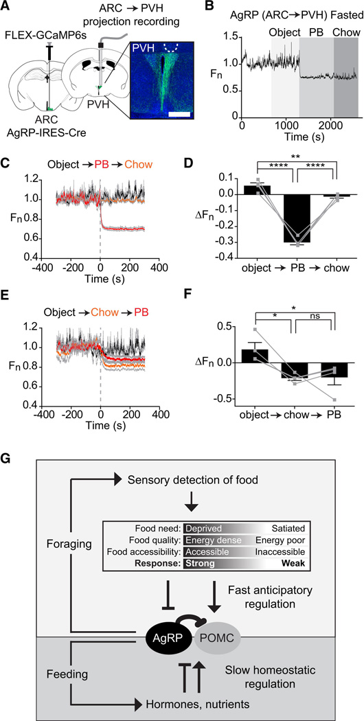
Hunger is controlled by specialized neural circuits that translate homeostatic needs into motivated behaviors. These circuits are under chronic control by circulating signals of nutritional state, but their rapid dynamics on the timescale of behavior remain unknown. Here, we report optical recording of the natural activity of two key cell types that control food intake, AgRP and POMC neurons, in awake behaving mice. We find unexpectedly that the sensory detection of food is sufficient to rapidly reverse the activation state of these neurons induced by energy deficit. This rapid regulation is cell-type specific, modulated by food palatability and nutritional state, and occurs before any food is consumed. These data reveal that AgRP and POMC neurons receive real-time information about the availability of food in the external world, suggesting a primary role for these neurons in controlling appetitive behaviors such as foraging that promote the discovery of food.


Figures

Figure 1
Figure 1. Optical recording of AgRP and POMC neuron activity in awake behaving mice
(A) FLEX AAV used to drive GCaMP6s expression. (B) Response of AgRP and POMC neurons to current ramp. Scale bar indicates GCaMP6s fluorescence normalized to 1.0 at start of the experiment (Fn). (C) Membrane potential and GCaMP6s fluorescence in response to sequential 10 pA current steps of duration 2s separated by 20s. (D) Relationship between action potential number and fluorescence for cells in panel C. (E) R-squared and p values for the linear regression of fluorescence versus action potential number for 16 POMC and 14 AgRP neurons. (F) Schematic of the fiber photometry setup. (G) Coronal section from AgRP and POMC mice showing path of optical fiber and injection site. Scale bar = 1 mm (H) Fluorescence trace during cage exploration for mice expressing GCaMP6s or GFP in AgRP neurons or POMC neurons. See also Figure S1.
Figure 2
Figure 2. Ghrelin rapidly modulates AgRP and POMC neurons
(A and C) Recordings from a mouse expressing GCaMP6s in AgRP or POMC neurons that was challenged with injection of ghrelin (light gray) followed by presentation of a pellet of chow (dark gray). (B and D). Calcium signals from AgRP and POMC neurons aligned to the time of PBS or ghrelin injection, or chow presentation to ghrelin treated mice. Red and gray indicate the mean response and standard error (AgRP, n=7; POMC, n=5). In each trial fluorescence was normalized by assigning a value of 1.0 to the median value of data points within a two minute window at −5 min before treatment. (E) Peri-event plots showing the response from a single trial of five AgRP mice and five POMC mice.
Figure 3
Figure 3. Sensory detection of food rapidly regulates AgRP and POMC neurons
(A and D) Recordings from fasted mice expressing GCaMP6s in AgRP or POMC neurons presented with a pellet of chow (gray). (B and E) Plots of calcium signals from AgRP and POMC neurons aligned to the time of presentation of a pellet of chow (red) or inedible object (black). Mice were either subjected to an overnight fast (left) or fed ad libitum (right) prior to experiment. Gray indicates standard error (AgRP, n=10; POMC, n=5). (C and F) Quantification of fluorescence changes 5 min after event, as indicated. (G) Peri-event plots aligned to the time of event. Each row is a single trial of a different mouse. (H) Calcium signals aligned to the initiation of feeding for AgRP and POMC neurons. (I) Quantification of change in fluorescence occurs before feeding is initiated versus the total change in the trial. * p<0.05. ** p<0.01,*** p<0.001,**** p<0.0001.
Figure 4
Figure 4. Food palatability determines the magnitude of the response to food detection
(A and C) Calcium signals from AgRP and POMC neurons in fasted and fed mice aligned to the time of presentation of peanut butter or chow. (B and D) Fluorescence change of AgRP and POMC neurons upon sequential presentation of an inedible object, chow, and peanut butter in fasted mice. (E) Quantification of responses of AgRP and POMC neurons 5 min after food presentation. (F) Plot showing the response of AgRP and POMC neurons over 5 min to different foods and pharmacologic treatments in the context of varying nutritional states. All traces start at the origin (0,0) and emanate outward. Arrows indicate the direction of movement. See also Figure S2.
Figure 5
Figure 5. The response to food detection depends on food accessibility and is reversible
(A) Schematic of caged peanut butter. (B) Calcium signals aligned to the time of presentation of a caged peanut butter. (C) Change in fluorescence in 1 and 8 min after caged peanut butter presentation. (D) Schematic of hidden peanut butter. (E) Calcium signals aligned to the time of presentation of a hidden peanut butter. (F) Change in fluorescence in 1 and 8 min after hidden peanut butter presentation. (G and J) Chow was presented at time 0, and then food was removed at 2 min (red), 10 min (blue) or not removed (black). (H and K) Recovery in fluorescence 20 min after food removal for experiments in which food was removed after 2, 10, or 30 min. (I and L) Time constant for the response to upon food presentation and food removal after 2 and 10 min. See also Figure S3.
Figure 6
Figure 6. Intrameal dynamics of AgRP and POMC neurons
(A, and B) Traces of AgRP and POMC activity in mice during consumption of a liquid diet. Licks that mark initiation of a feeding bout are shown in gray. (C) Difference in average fluorescence between periods of feeding (intrabout) and intermeal intervals (interbout) for each mouse. (D, E) Calcium signals from AgRP and POMC neurons aligned to the moment of the first lick that initiates a feeding bout. Data from actual feeding bouts shown in red; data from simulated randomly generated feeding bouts in black. (F) Cross-correlation plots showing the correlation between activity of AgRP and POMC neurons before and after licking. Red is mean, gray is 28 individual comparisons between AgRP (n=7) and POMC (n=4) mice. (G and H) Peri-event plots showing the activity of AgRP and POMC neurons aligned to the start of feeding bouts. The top plot shows all of the bouts for one trial of a mouse. The bottom plot shows the average response across all bouts for 7 AgRP and 4 POMC mice.
Figure 7
Figure 7. Natural dynamics of AgRP projections to the PVH
(A) Schematic showing infection of cell bodies in the ARC and installation of optical fiber in the PVH. Scale bar = 0.5 mm (B) Recording from PVH of a fasted mouse presented sequentially with an inedible object, peanut butter, and chow. (C and E) Calcium signals from from PVH of mice presented sequentially with an inedible object, peanut butter, and chow. (D and F) Quantification of calcium signals five minutes after event. (n=4 mice). (G) Model for regulation of AgRP and POMC neurons by homeostatic and sensory information. See also Figure S4.

Comment in

  • The hunger games.
    Seeley RJ, Berridge KC. Seeley RJ, et al. Cell. 2015 Feb 26;160(5):805-806. doi: 10.1016/j.cell.2015.02.028. Cell. 2015. PMID: 25723156

Similar articles

Cited by

References

    1. Aponte Y, Atasoy D, Sternson SM. AGRP neurons are sufficient to orchestrate feeding behavior rapidly and without training. Nature neuroscience. 2011;14:351–355. - PMC - PubMed
    1. Atasoy D, Betley JN, Su HH, Sternson SM. Deconstruction of a neural circuit for hunger. Nature. 2012;488:172–177. - PMC - PubMed
    1. Betley JN, Cao ZF, Ritola KD, Sternson SM. Parallel, redundant circuit organization for homeostatic control of feeding behavior. Cell. 2013;155:1337–1350. - PMC - PubMed
    1. Blouet C, Schwartz GJ. Hypothalamic nutrient sensing in the control of energy homeostasis. Behavioural brain research. 2010;209:1–12. - PubMed
    1. Chen TW, Wardill TJ, Sun Y, Pulver SR, Renninger SL, Baohan A, Schreiter ER, Kerr RA, Orger MB, Jayaraman V, et al. Ultrasensitive fluorescent proteins for imaging neuronal activity. Nature. 2013;499:295–300. - PMC - PubMed

Publication types

Substances