

Sensitization of enteric neurons to morphine by HIV-1 Tat protein
- PMID: 25703354
- PMCID: PMC4380805
- DOI: 10.1111/nmo.12514
Sensitization of enteric neurons to morphine by HIV-1 Tat protein
Abstract
Background: Gastrointestinal (GI) dysfunction is a major cause of morbidity in acquired immunodeficiency syndrome (AIDS). HIV-1-induced neuropathogenesis is significantly enhanced by opiate abuse, which increases proinflammatory chemokine/cytokine release, the production of reactive species, glial reactivity, and neuronal injury in the central nervous system. Despite marked interactions in the gut, little is known about the effects of HIV-1 in combination with opiate use on the enteric nervous system.
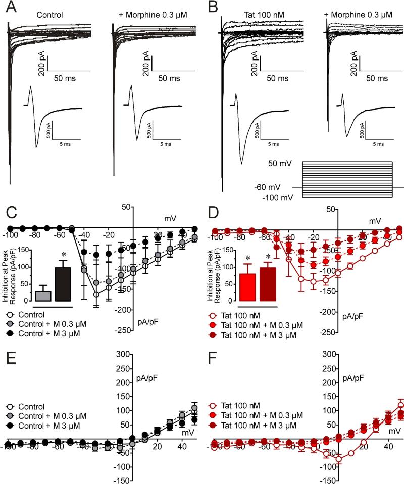
Methods: To explore HIV-opiate interactions in myenteric neurons, the effects of Tat ± morphine (0.03, 0.3, and 3 μM) were examined in isolated neurons from doxycycline- (DOX-) inducible HIV-1 Tat(1-86) transgenic mice or following in vitro Tat 100 nM exposure (>6 h).
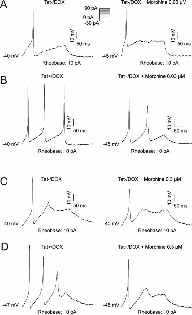
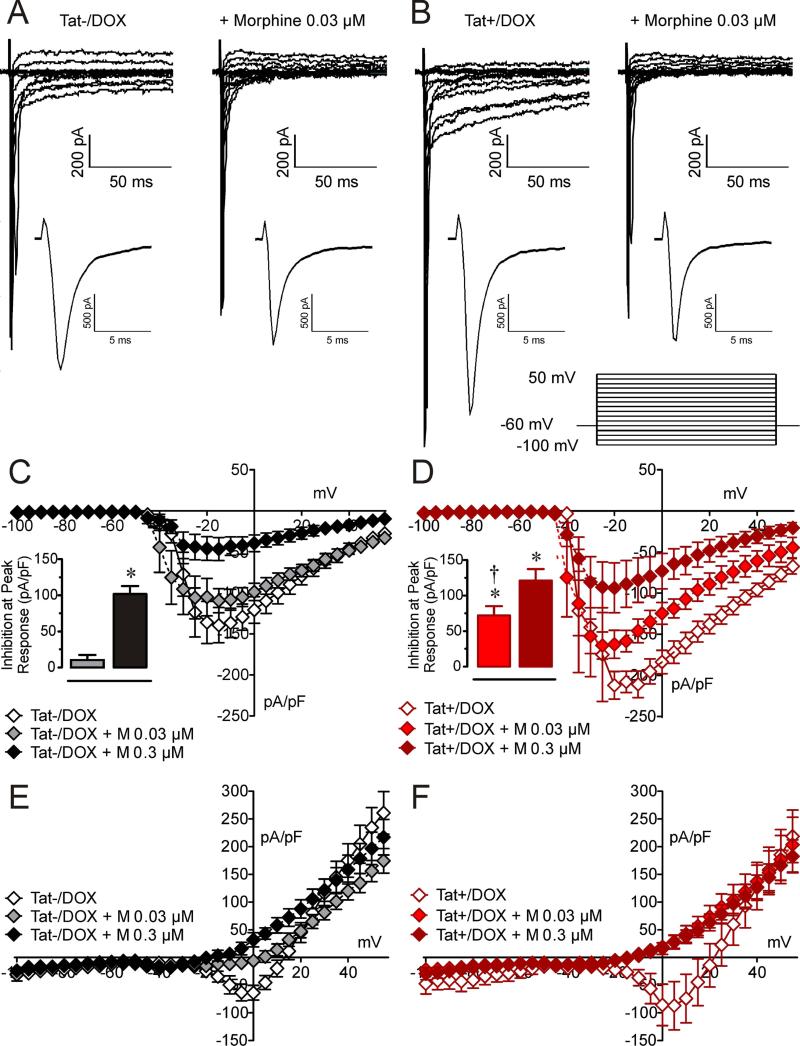
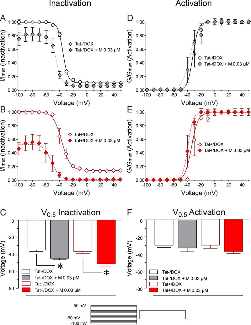
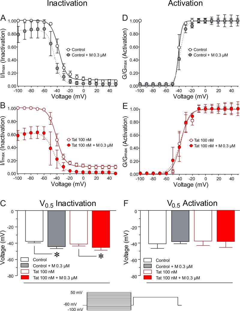
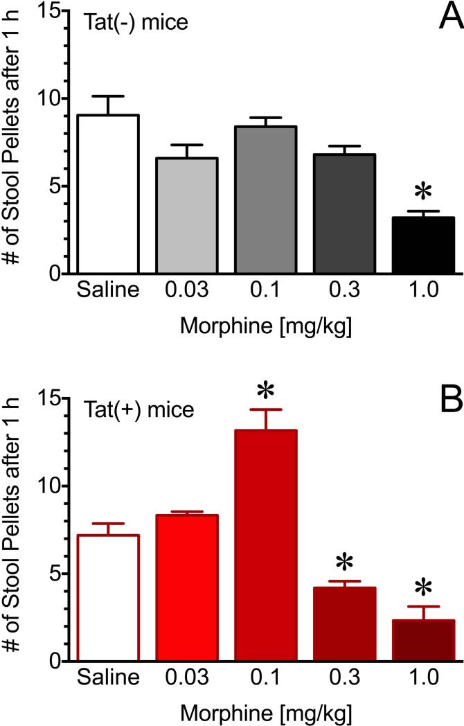
Key results: Current clamp recordings demonstrated increased neuronal excitability in neurons of inducible Tat(+) mice (Tat+/DOX) compared to control Tat-/DOX mice. In neurons from Tat+/DOX, but not from Tat-/DOX mice, 0.03 μM morphine significantly reduced neuronal excitability, fast transient and late long-lasting sodium currents. There was a significant leftward shift in V(0.5) of inactivation following exposure to 0.03 μM morphine, with a 50% decrease in availability of sodium channels at -100 mV. Similar effects were noted with in vitro Tat exposure in the presence of 0.3 μM morphine. Additionally, GI motility was significantly more sensitive to morphine in Tat(+) mice than Tat(-) mice.
Conclusions & inferences: Overall, these data suggest that the sensitivity of enteric neurons to morphine is enhanced in the presence of Tat. Opiates and HIV-1 may uniquely interact to exacerbate the deleterious effects of HIV-1-infection and opiate exposure on GI function.
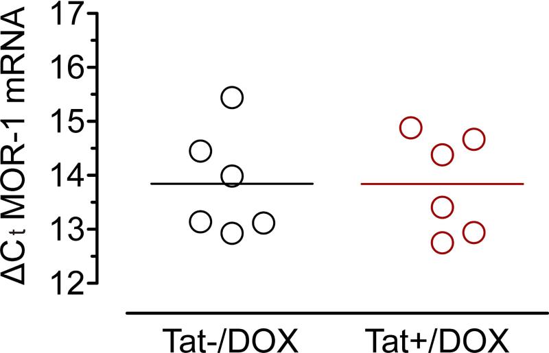
Keywords: HIV-1 Tat; MOR-1 expression; enteric neurons; excitability; gut; ileum; opioid drug abuse; sodium currents.
© 2015 John Wiley & Sons Ltd.
Figures
Similar articles
-
Effects of HIV-1 Tat on enteric neuropathogenesis.J Neurosci. 2014 Oct 22;34(43):14243-51. doi: 10.1523/JNEUROSCI.2283-14.2014. J Neurosci. 2014. PMID: 25339738 Free PMC article.
-
HIV, opiates, and enteric neuron dysfunction.Neurogastroenterol Motil. 2015 Apr;27(4):449-54. doi: 10.1111/nmo.12539. Neurogastroenterol Motil. 2015. PMID: 25817054 Free PMC article. Review.
-
A central role for glial CCR5 in directing the neuropathological interactions of HIV-1 Tat and opiates.J Neuroinflammation. 2018 Oct 10;15(1):285. doi: 10.1186/s12974-018-1320-4. J Neuroinflammation. 2018. PMID: 30305110 Free PMC article.
-
Central HIV-1 Tat exposure elevates anxiety and fear conditioned responses of male mice concurrent with altered mu-opioid receptor-mediated G-protein activation and β-arrestin 2 activity in the forebrain.Neurobiol Dis. 2016 Aug;92(Pt B):124-36. doi: 10.1016/j.nbd.2016.01.014. Epub 2016 Feb 1. Neurobiol Dis. 2016. PMID: 26845176 Free PMC article.
-
Opiate drug use and the pathophysiology of neuroAIDS.Curr HIV Res. 2012 Jul;10(5):435-52. doi: 10.2174/157016212802138779. Curr HIV Res. 2012. PMID: 22591368 Free PMC article. Review.
Cited by
-
HIV-1 Tat-induced diarrhea evokes an enteric glia-dependent neuroinflammatory response in the central nervous system.Sci Rep. 2017 Aug 10;7(1):7735. doi: 10.1038/s41598-017-05245-9. Sci Rep. 2017. PMID: 28798420 Free PMC article.
-
Inhibition of GABAergic Neurotransmission by HIV-1 Tat and Opioid Treatment in the Striatum Involves μ-Opioid Receptors.Front Neurosci. 2016 Nov 8;10:497. doi: 10.3389/fnins.2016.00497. eCollection 2016. Front Neurosci. 2016. PMID: 27877102 Free PMC article.
-
Mechanisms of enteric neuropathy in diverse contexts of gastrointestinal dysfunction.Neurogastroenterol Motil. 2025 Aug;37(8):e14870. doi: 10.1111/nmo.14870. Epub 2024 Jul 22. Neurogastroenterol Motil. 2025. PMID: 39038157 Free PMC article. Review.
-
The gut-brain interaction in opioid tolerance.Curr Opin Pharmacol. 2017 Dec;37:126-130. doi: 10.1016/j.coph.2017.10.012. Epub 2017 Nov 13. Curr Opin Pharmacol. 2017. PMID: 29145012 Free PMC article. Review.
-
HIV-1 Tat-induced diarrhea is improved by the PPARalpha agonist, palmitoylethanolamide, by suppressing the activation of enteric glia.J Neuroinflammation. 2018 Mar 24;15(1):94. doi: 10.1186/s12974-018-1126-4. J Neuroinflammation. 2018. PMID: 29573741 Free PMC article.
References
-
- Anthony IC, Arango JC, Stephens B, Simmonds P, Bell JE. The effects of illicit drugs on the HIV infected brain. Front Biosci. 2008;13:1294–1307. - PubMed
-
- Anthony IC, Ramage SN, Carnie FW, Simmonds P, Bell JE. Does drug abuse alter microglial phenotype and cell turnover in the context of advancing HIV infection? Neuropathol Appl Neurobiol. 2005;31:325–338. - PubMed
-
- Bell JE, Brettle RP, Chiswick A, Simmonds P. HIV encephalitis, proviral load and dementia in drug users and homosexuals with AIDS. Effect of neocortical involvement. Brain. 1998;121(Pt 11):2043–2052. - PubMed
-
- Bell JE, Arango JC, Anthony IC. Neurobiology of multiple insults: HIV-1-associated brain disorders in those who use illicit drugs. J Neuroimmune Pharmacol. 2006;1:182–191. - PubMed
-
- Wilcox CM, Rabeneck L, Friedman S. AGA technical review: malnutrition and cachexia, chronic diarrhea, and hepatobiliary disease in patients with human immunodeficiency virus infection. Gastroenterology. 1996;111:1724–1752. - PubMed
Publication types
MeSH terms
Substances
Grants and funding
LinkOut - more resources
Full Text Sources
Other Literature Sources
Molecular Biology Databases
