. 2015 Mar 3;23(3):517-526.
doi: 10.1016/j.str.2015.01.012. Epub 2015 Feb 19.

A conserved activation cluster is required for allosteric communication in HtrA-family proteases

Affiliations

A conserved activation cluster is required for allosteric communication in HtrA-family proteases

Anna K de Regt et al. Structure. .

Abstract

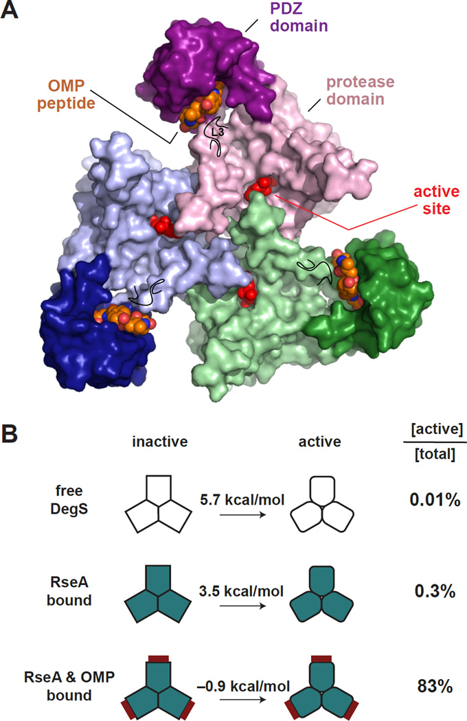
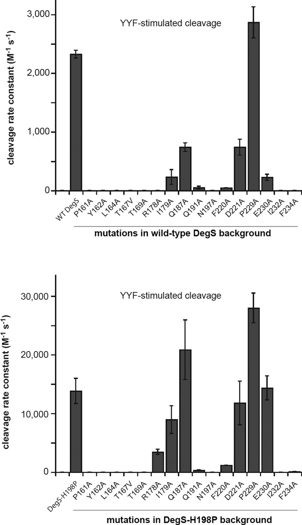
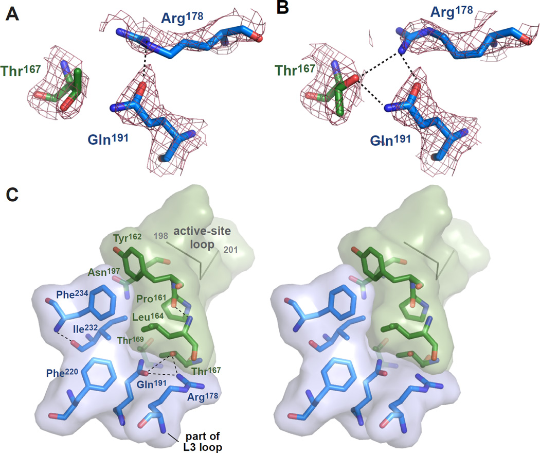
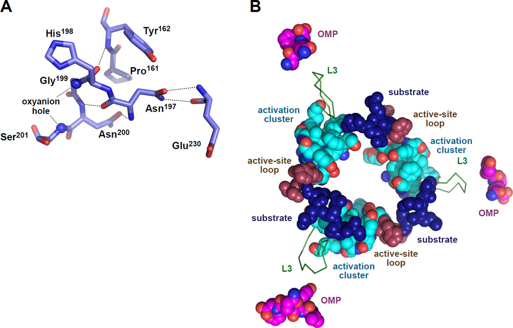
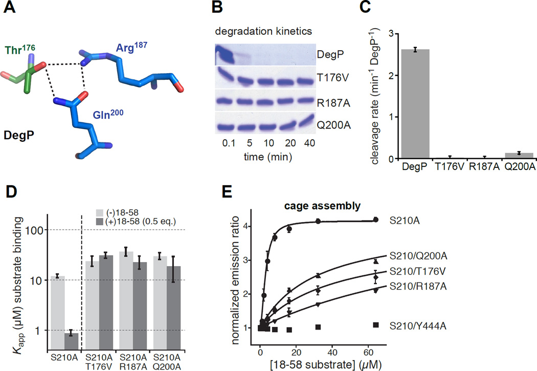
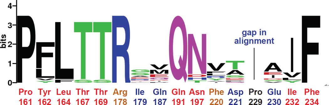
In E. coli, outer-membrane stress causes a transcriptional response through a signaling cascade initiated by DegS cleavage of a transmembrane antisigma factor. Each subunit of DegS, an HtrA-family protease, contains a protease domain and a PDZ domain. The trimeric protease domain is autoinhibited by the unliganded PDZ domains. Allosteric activation requires binding of unassembled outer-membrane proteins (OMPs) to the PDZ domains and protein substrate binding. Here, we identify a set of DegS residues that cluster together at subunit-subunit interfaces in the trimer, link the active sites and substrate binding sites, and are crucial for stabilizing the active enzyme conformation in response to OMP signaling. These residues are conserved across the HtrA-protease family, including orthologs linked to human disease, supporting a common mechanism of allosteric activation. Indeed, mutation of residues at homologous positions in the DegP quality-control protease also eliminates allosteric activation.


Figures

Figure 1
Figure 1. The DegS Trimer Equilibrates Between Inactive and Active Structures
(A) Structure of a DegS trimer in the active conformation with OMP peptides bound to the PDZ domains (pdb code 3GDS). Each DegS subunit (surface representation colored green, purple, or blue) consists of a protease domain (lighter colors), which pack together to form the trimer, and a peripheral PDZ domain (darker colors). Bound OMP peptides are shown in CPK representation with orange carbons. An isopropyl phosphate group covalently bound to the active-site serine is shown in CPK representation and colored red. The position of the L3 loop is marked by a black line. (B) Free energies for conversion of inactive to active DegS in the free enzyme, the substrate-saturated enzyme, and the substrate and OMP-peptide saturated enzyme (Sohn and Sauer, 2009). These values would be decreased by ~4 kcal/mol for H198P DegS (Sohn et al., 2009; 2010). The column on the right lists the percentage of total DegS that assumes the active conformation for the unbound and ligand bound states.
Figure 2
Figure 2. Mutational Effects on DegS Cleavage of RseA
Effects of DegS mutations in an otherwise wild-type background (upper panel) or in an H198P background (lower panel) on OMP-peptide stimulated proteolytic activity, expressed as the second-order rate constant (kcat/KM) for RseA cleavage. Values are averages of two or more independent trials ± SEM. Cleavage reactions contained different sub-KM concentrations of 35S-labelled RseA, DegS or variants (1 µM trimer), and YYF OMP peptide (230 µM). Initial cleavage rates normalized by total enzyme were plotted as a function of the RseA concentration, and the second-order rate constant was determined from the slope of a linear fit.
Figure 3
Figure 3. Structural Determinants of DegS Activation
(A) Electron-density map (2FO–FC; contoured at 1.3 σ) for Thr167 in chain B (dark green carbons) and Arg178 and Gln191 in chain C (marine carbons) in the 1SOZ DegS trimer. Thr167 is modeled in an eclipsed conformation (χ 116°) found in only 0.1% of high-resolution structures. A hydrogen bond is shown as a dashed black line. (B) Electron-density map (same residues and parameters as panel A) but for the 4RQZ re-refined structure of 1SOZ. Thr167 is modeled as a rotamer (χ 290°) found in 18.7% of high-resolution structures and forms hydrogen bonds (dashed black lines) with Arg178 and Gln191. (C) Stereo view of residues identified as being very important or somewhat important for DegS allostery cluster between the active-site loop containing the catalytic Ser201 and oxyanion hole (top right) and the L3 loop (bottom center). Residues with marine-colored carbons are from one subunit of the 4RQZ trimer and those with dark-green carbons are from another subunit; transparent surfaces are shown for both subunits to emphasize the position of the interface. The residues shown represent one of the three activation clusters in the DegS trimer. Hydrogen bonds are represented as dashed black lines.
Figure 4
Figure 4. Allosteric Ligands and Catalytic and Regulatory Elements in Active DegS
(A) Main-chain and side-chain atoms from activation-cluster residues Pro161, Tyr162, and Asn197 stabilize the functional conformation of the active-site loop, including the catalytic Ser201 and the oxyanion-hole nitrogens (small spheres). Hydrogen bonds are shown as dashed lines. (B) Positions of OMP peptides, L3 loops, activation clusters, active-site loops, and modeled substrate in an active DegS trimer (4RQZ). The position of substrate was modeled by aligning 4RQZ with a DegS ortholog from Mycobacterium tuberculosis with a bound substrate-cleavage product (2Z9I; Mohamedmohaideen et al., 2008). Although the PDZ-bound OMP peptides are adjacent to the L3 loops, there are no critical residues between the OMP peptide and the activation clusters, suggesting that OMP-peptide binding destabilizes autoinhibitory interactions rather than directly stabilizing the active conformation. Substrate binding ties together all of the active-site loops and activation clusters in the trimer, providing a mechanism for allosteric substrate stabilization of the active conformation.
Figure 5
Figure 5. Effects of activation-cluster mutations on DegP activity and cage assembly
(A) The side chain of Thr176 from one DegP subunit (dark-green carbons) forms hydrogen bonds with the side chains of Arg187 and Gln200 in an adjacent subunit (marine carbons) in the protease domain of an active DegP trimer (3OTP). (B) Kinetics of degradation of carboxymethylated lysozyme (5 µM) by wild-type DegP and the T176V, R187A, or Q200A variants (1 µM) at room temperature was assayed by SDS-PAGE and staining with Coomassie Blue. (C) Rates of cleavage of the p23 substrate (40 µM) by wild-type DegP or variants (1 µM) were determined at room temperature. Error bars are averages ±1 SD (N = 3). (D) Apparent equilibrium dissociation constants (Kapp) for binding of different concentrations of proteolytically inactive DegPS210A or variants to a fluorescent substrate (flClys18–58; 50 nM) were determined in the absence or presence of unlabeled lys18–58 at one-half of the DegP concentration by changes in fluorescence anisotropy (Kim et al., 2011; Kim and Sauer, 2012). Positive cooperativity makes binding stronger in the presence of unlabeled lys18–58 for variants, like DegPS210A, that can switch between the inactive and active conformations. The T167V, R187A, and Q200A mutations weakened binding and eliminated positive cooperativity. Bars represent the error of fitting of individual binding curves to a hyperbolic binding equation. (E) Polyhedral-cage assembly of DegP variants as a function of added lys18–58 substrate was monitored by increases in FRET between trimers labeled with donor and acceptor dyes (Kim et al., 2011). Emission ratios were normalized to data in the absence of substrate and fitted to a Hill equation (y = y0 + max • [lys18–58]h/(Kapph + [lys18–58]h)) for DegPS210A or to a hyperbolic equation (y = y0 + max • [lys18–58]/(Kapp + [lys18–58])) for variants containing the T176V, R187A, or Q200A mutations. No assembly of the DegPS210A/Y444A variant was observed, as expected because the Y444A mutation removes a critical interaction that stabilizes cages (Kim et al., 2011). Error bars are averages ±1 SD (N = 3).
Figure 6
Figure 6. Side Chains Important for DegS Allostery Are Conserved in the HtrA Family
Residues shown here to be very important (red labels) or somewhat important (orange labels) for allosteric activation of DegS are largely conserved in HtrA proteases from all kingdoms of life. Residues that play minor roles (blue labels) or no role (black label) in DegS activity are far less conserved. HtrA sequences from E. coli (3), H. sapiens (4), Synechocystis (3), D. rerio, A. thaliana, C. reinhardtii, D. melanogaster, B. dendrobatidis, A. pseudotrichonymphae, and K. cryptofilum were aligned using ClustalW2 (Larkin et al., 2007) and submitted to WebLogo (Crooks et al., 2004) for visualization of conservation information. A higher number of bits indicates greater conservation for a given position.

Similar articles

Cited by

References

    1. Adams PD, et al. PHENIX: a comprehensive Python-based system for macromolecular structure solution. Acta Crystallogr. D. 2010;66:213–221. - PMC - PubMed
    1. Ades SE, Connolly LE, Alba BM, Gross CA. The Escherichia coli sigma(E)-dependent extracytoplasmic stress response is controlled by the regulated proteolysis of an anti-sigma factor. Genes Dev. 1999;13:2449–2461. - PMC - PubMed
    1. Ades SE. Regulation by destruction: design of the σE envelope stress response. Curr. Opin. Microbiol. 2008;11:535–540. - PubMed
    1. Akiyama Y, Kanehara K, Ito K. RseP (YaeL), an Escherichia coli RIP protease, cleaves transmembrane sequences. EMBO J. 2004;23:4434–4442. - PMC - PubMed
    1. Alba BM, Gross CA. Regulation of the Escherichia coli sigma-dependent envelope stress response. Mol. Microbiol. 2004;52:613–619. - PubMed

Publication types

LinkOut - more resources