Review
. 2015 Mar;264(1):182-203.
doi: 10.1111/imr.12266.

Macrophage defense mechanisms against intracellular bacteria

Affiliations
Review

Macrophage defense mechanisms against intracellular bacteria

Günter Weiss et al. Immunol Rev. 2015 Mar.

Abstract

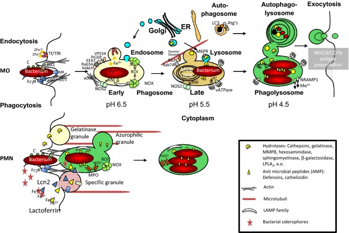
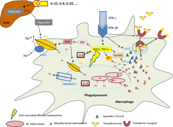
Macrophages and neutrophils play a decisive role in host responses to intracellular bacteria including the agent of tuberculosis (TB), Mycobacterium tuberculosis as they represent the forefront of innate immune defense against bacterial invaders. At the same time, these phagocytes are also primary targets of intracellular bacteria to be abused as host cells. Their efficacy to contain and eliminate intracellular M. tuberculosis decides whether a patient initially becomes infected or not. However, when the infection becomes chronic or even latent (as in the case of TB) despite development of specific immune activation, phagocytes have also important effector functions. Macrophages have evolved a myriad of defense strategies to combat infection with intracellular bacteria such as M. tuberculosis. These include induction of toxic anti-microbial effectors such as nitric oxide and reactive oxygen intermediates, the stimulation of microbe intoxication mechanisms via acidification or metal accumulation in the phagolysosome, the restriction of the microbe's access to essential nutrients such as iron, fatty acids, or amino acids, the production of anti-microbial peptides and cytokines, along with induction of autophagy and efferocytosis to eliminate the pathogen. On the other hand, M. tuberculosis, as a prime example of a well-adapted facultative intracellular bacterium, has learned during evolution to counter-balance the host's immune defense strategies to secure survival or multiplication within this otherwise hostile environment. This review provides an overview of innate immune defense of macrophages directed against intracellular bacteria with a focus on M. tuberculosis. Gaining more insights and knowledge into this complex network of host-pathogen interaction will identify novel target sites of intervention to successfully clear infection at a time of rapidly emerging multi-resistance of M. tuberculosis against conventional antibiotics.

Keywords: efferocytosis; iron; macrophages; phagolysosome; radicals; vitamin D.


Figures

Figure 1
Figure 1
Intracellular trafficking of bacteria in phagocytes and anti-microbial responses in macrophages and polymorphonuclear neutrophils. Schematic drawing depicts the phagocytic responses of macrophages (MΦs) and neutrophils (PMNs) against bacterial invaders, and the intracellular fate of the engulfed particles. Non-opsonized or antibody-/complement (C)-opsonized bacteria are recognized and bound by surface receptors for bacterial compounds or the respective opsonins, which triggers signaling cascades involving Syk or DIA1 and leads to actin polymerization and phagocytic cup formation. In MΦs, upon phagosome closure, the maturing phagosome traverses an early and late phagosomal and a phagolysosomal stage paralleling endosomal maturation. Phagosome biogenesis is accompanied by continuous fusion and fission events, including fusion with trans-Golgi transport vesicles, endosomes, lysosomes, and autophagosomes. These interactions cause acquisition and loss of different stage-specific markers. A hallmark of phagosome biogenesis is acidification of the phagosomal lumen by the proton pumping vATPase. A low pH is a prerequisite for optimal enzymatic activity of most late endosomal/lysosomal hydrolases, which are delivered to the nascent phagosome bound to the M6PR from the trans-Golgi. The stepwise succession of phagosomal maturation in macrophages is strikingly different from phagosome formation in PMNs. Phagocytosis and fusion with the lysosomal azurophilic granules is often happening simultaneously. At the same time, specific granules discharge iron sequestering lactoferrin and Lcn2 into both, the phagosomal lumen and the extracellular space. These granules also deliver phagocytic receptors to the PMN surface for recognition and uptake of bacteria. Finally, the gelatinase granules spill out proteases and other enzymes to degrade extracellular matrix proteins, leading to tissue disruption to allow PMN evasion into infection site but ultimately to pathogenesis.
Figure 2
Figure 2
The battle for iron. Macrophages use multiple pathways to restrict the essential growth factor iron for intracellular mycobacteria. First, cytokines such as IFN-γ inhibit the transcriptional expression of transferrin receptor (TfR). TfR is a major source of iron for mycobacteria, because the bacteria can utilize its ligand, transferrin iron, following its endosomal transfer. Macrophages produce lipocalin-2 (Lcn2), which binds and neutralizes siderophores produced by M. tuberculosis to scavenge and re-utilize cytoplasmic iron. Furthermore, macrophage-derived cytokines such as TNF-α induce the formation of the iron-binding protein ferritin, which incorporates iron into its core rendering it unavailable for intracellular bacteria as well as the iron binding protein lactoferrin (Lf), which also scavenges this metal. Activated macrophages express the phagolysomal protein Nramp1, which among other effects pumps iron out of macrophages, thereby reducing the availability of the metal in the phagosome for mycobacteria. Finally, upon formation of nitric oxide, the transcription factor Nrf2 is activated, stimulating the expression of the major iron export protein ferroportin (FP1) pumping iron out of the phagolyosome and of the cytoplasma of macrophages. By mechanisms that remain elusive, the stimulation of hepcidin expression in the liver, which is a major mechanism for iron restriction to extracellular pathogens, is circumvented. All these events result in reduction in intracellular iron levels and a limited availability of iron for intra-macrophage bacteria. Based on the negative regulatory effects of iron on IFN-γ activity, the reduction in this metal's availability results in strengthened innate anti-microbial immune responses. Importantly, some of the pathways shown in this figure have been investigated for other intracellular bacteria such as S. typhimurium, and their importance for M. tuberculosis remains to be shown.

Similar articles

Cited by

References

    1. Watson RO, Manzanillo PS, Cox JS. Extracellular M. tuberculosis DNA targets bacteria for autophagy by activating the host DNA-sensing pathway. Cell. 2012;150:803–815. - PMC - PubMed
    1. Sun L, Wu J, Du F, Chen X, Chen ZJ. Cyclic GMP-AMP synthase is a cytosolic DNA sensor that activates the type I interferon pathway. Science. 2013;339:786–791. - PMC - PubMed
    1. Sambarey A, Prashanthi K, Chandra N. Mining large-scale response networks reveals ‘topmost activities’ in Mycobacterium tuberculosis infection. Sci Rep. 2013;3:2302. - PMC - PubMed
    1. Bruns H, Stenger S. New insights into the interaction of Mycobacterium tuberculosis and human macrophages. Future Microbiol. 2014;9:327–341. - PubMed
    1. Schnappinger D, et al. Transcriptional adaptation of Mycobacterium tuberculosis within macrophages: insights into the phagosomal environment. J Exp Med. 2003;198:693–704. - PMC - PubMed

Publication types

MeSH terms