Review
. 2015 Mar;264(1):288-307.
doi: 10.1111/imr.12252.

Heterogeneity in tuberculosis pathology, microenvironments and therapeutic responses

Affiliations
Review

Heterogeneity in tuberculosis pathology, microenvironments and therapeutic responses

Anne Lenaerts et al. Immunol Rev. 2015 Mar.

Abstract

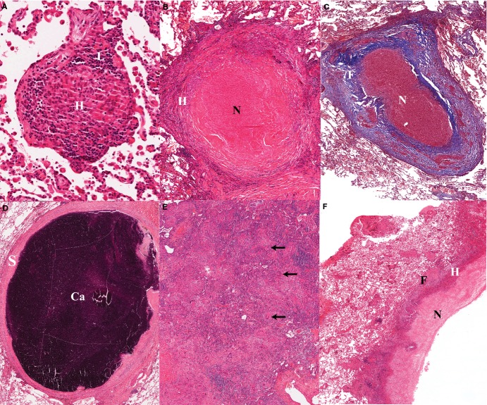
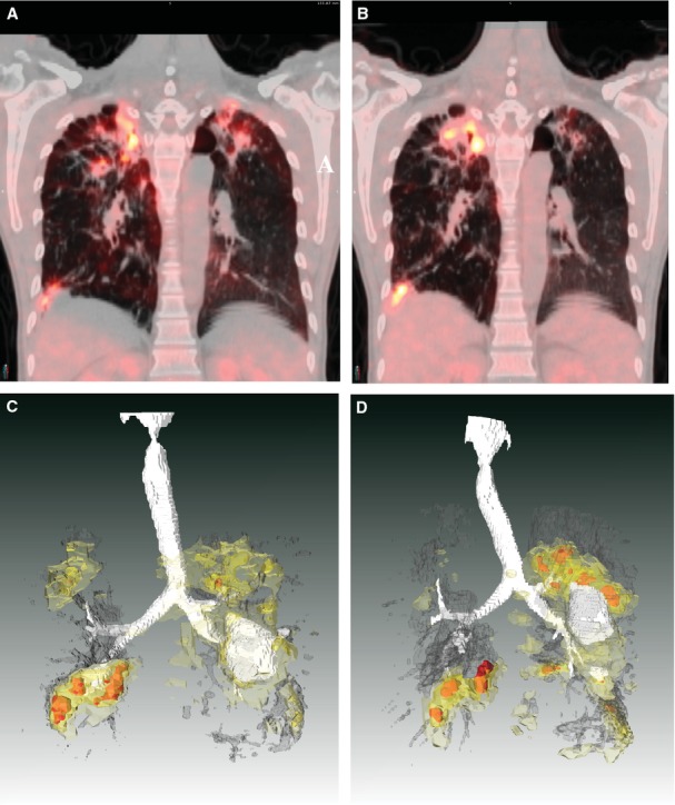
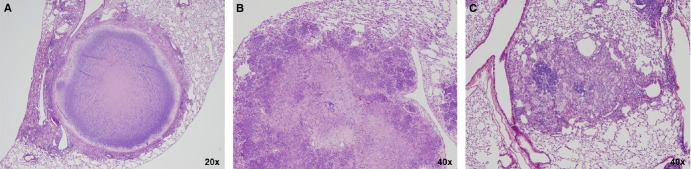
Tuberculosis (TB) lesions are extremely complex and dynamic. Here, we review the multiple types and fates of pulmonary lesions that form following infection by Mycobacterium tuberculosis and the impact of this spatial and temporal heterogeneity on the bacteria they harbor. The diverse immunopathology of granulomas and cavities generates a plethora of microenvironments to which M. tuberculosis bacilli must adapt. This in turn affects the replication, metabolism, and relative density of bacterial subpopulations, and consequently their respective susceptibility to chemotherapy. We outline recent developments that support a paradigm shift in our understanding of lesion progression. The simple model according to which lesions within a single individual react similarly to the systemic immune response no longer prevails. Host-pathogen interactions within lesions are a dynamic process, driven by subtle and local differences in signaling pathways, resulting in diverging trajectories of lesions within a single host. The spectrum of TB lesions is a continuum with a large overlap in the lesion types found in latently infected and active TB patients. We hope this overview will guide TB researchers in the design, choice of read-outs, and interpretation of future studies in the search for predictive biomarkers and novel therapies.

Keywords: animal models; biomarkers; immunopathology; lesion dynamics; tuberculosis.


Figures

Figure 1
Figure 1
(A). Early non-necrotizing granuloma; the lymphocyte rim (L) appears bluish owing to the prominent nuclei and small cytoplasm; the center of the granuloma contains a majority of epitheloid histiocytes (H) or macrophages, which are large cells with pale abundant cytoplasm. (B). Necrotizing granuloma: the central necrosis appears as pink amorphous material (N), surrounded by a layer of cellular infiltrates as well as a fibrotic rim (F) which clearly defines the granuloma from the surrounding lung alveoli. (C). Masson's trichrome stain of a necrotizing granuloma showing the collagenous areas in blue. The thick fibrotic layer (F) is composed of fibroblasts, collagen and new vessels, and scattered chronic inflammatory cells. Note the prominent vascularization of the fibrotic layer. (D). Calcified granuloma with sclerotic rim (S) associated with absence of mononuclear infiltrates and no visible infectious activity; Ca: area of calcification/mineralization. (E). Tuberculous pneumonia: confluent aggregates of epitheloid histiocytes mixed with Langhans’ and multinuclear giant cells (arrows). The more bluish areas are lymphoid aggregates. These are the same cells that make up a granuloma, but in the absence of a fibrotic wall they have spread to fill alveolar space. (F). Wall of cavitating tuberculous granuloma demonstrating a trilaminar structure with inner necrosis (N), histiocyte aggregates (H) and peripheral fibrosis (F). The fibrotic wall contains mononuclear infiltrates which indicates a still active and progressing tuberculous lesion. The more friable caseous material has been removed during tissue processing.
Figure 2
Figure 2
TB lesions evolve independently in humans. This patient had non-responsive extensively drug-resistant TB and was enrolled into the delayed arm of a randomized clinical trial of linezolid. The subject had failed to respond to any chemotherapy and was hospitalized for 2 months while awaiting study drug. Upon admission, his baseline (18F)-2-fluorodeoxyglucose positron emission tomography (FDG-PET) computed tomography (CT) scan showed extensive bilateral disease. A coronal section of this subjects’ scan is shown in (A), projected at a maximum SUV (Standardized Uptake Value, a method to standardize FDG uptake accounting for body weight and decay time) of 10. The same coronal slice at the same SUV is shown from a repeated scan after 2 months (immediately prior to starting linezolid therapy) in (B). Apical lesions in the left lung (right side of PET/CT image) are resolving while apical lesions in the right lung have progressed. (C, D) The same scan data are presented from the full 3D volume of the chest cavity. White represents air, either in the trachea and airways or inside of cavitary lesions. Dark gray represents areas of radiodensity between −100 and 200 Hounsfeld Units associated with TB lesions in both lungs. FDG uptake is represented by light yellow (SUV 2-3) darker yellow (SUV 3-5) and red (SUV >5). These are posterior views, reversed from above for clarity. Each lesion complex appears to progress or regress independently of the others. The complex in the left superior lobe (on the left in these drawings) appears to resolve while the lesions in the right upper and lower lobes appear to progress significantly. The lesion in the left inferior lobe appears relatively stable with little change.
Figure 3
Figure 3
Heterogeneity in lesion types observed in C3HeB/FeJ mice infected with M. tuberculosis Erdman. Type I lesions (A) in C3HeB/FeJ mice are defined as the caseous necrotic lesions composed of a neutrophil-dominated central core region that degenerates over time into an amorphous, acellular caseum surrounded by a band of intact neutrophils and a distinct rim of foamy macrophages at the peripheral margin. The foamy macrophages contain numerous intracellular bacilli, while large numbers of extracellular bacilli are dispersed within the acellular caseum. The core region is encapsulated by a collagen rim deposited by fibroblasts intermixed with epithelioid and activated macrophages, and few lymphocytes. Type II lesions (B) observed in C3HeB/FeJ mice closely resemble PMN alveolitis occasionally observed in human tuberculosis patients, as described by Canetti (2). These lesions are predominantly composed of necrotizing neutrophils, but lack the fibrotic encapsulation seen in the Type I lesions. Type II lesions present as fulminant granulocytic pneumonia, containing high bacterial numbers extracellular and intracellular within in neutrophils, and very few if any detectable lymphocytes, and negatively impact mouse survival. Type III lesions (C) in C3HeB/FeJ mice are similar to lesions observed in BALB/c mice following aerosol infection. These inflammatory lesions are composed predominantly of epithelioid and foamy macrophages with large numbers of lymphocytes present, only containing few bacteria which are primarily located within macrophages.

Similar articles

Cited by

References

    1. Barnes DS. The Making of a Social Disease – Tuberculosis in Nineteenth-Century France. Berkeley, Los Angeles, London: University of California Press; 1995.
    1. Canetti G. The Tubercle Bacillus. New York: Springer Publishing COmpany; 1955.
    1. Dannenberg AM., Jr . Pathogenesis of Human Pulmonary Tuberculosis. Washington, DC: ASM Press; 2006.
    1. Via LE, et al. Infection dynamics and response to chemotherapy in a Rabbit model of tuberculosis using (18F)2-fluoro-deoxy-D-glucose positron emission tomography and computed tomography. Antimicrob Agents Chemother. 2012;56:4391–4402. - PMC - PubMed
    1. Via LE, et al. Differential virulence and disease progression following Mycobacterium tuberculosis complex infection of the common marmoset (Callithrix jacchus) Infect Immun. 2013;81:2909–2919. - PMC - PubMed

Publication types

Substances