. 2015 Mar 11;17(3):309-319.
doi: 10.1016/j.chom.2015.01.005. Epub 2015 Feb 19.

Influenza virus adaptation PB2-627K modulates nucleocapsid inhibition by the pathogen sensor RIG-I

Affiliations

Influenza virus adaptation PB2-627K modulates nucleocapsid inhibition by the pathogen sensor RIG-I

Michaela Weber et al. Cell Host Microbe. .

Abstract

The cytoplasmic RNA helicase RIG-I mediates innate sensing of RNA viruses. The genomes of influenza A virus (FLUAV) are encapsidated by the nucleoprotein and associated with RNA polymerase, posing potential barriers to RIG-I sensing. We show that RIG-I recognizes the 5'-triphosphorylated dsRNA on FLUAV nucleocapsids but that polymorphisms at position 627 of the viral polymerase subunit PB2 modulate RIG-I sensing. Compared to mammalian-adapted PB2-627K, avian FLUAV nucleocapsids possessing PB2-627E are prone to increased RIG-I recognition, and RIG-I-deficiency partially restores PB2-627E virus infection of mammalian cells. Heightened RIG-I sensing of PB2-627E nucleocapsids correlates with previously established lower affinity of 627E-containing PB2 for nucleoprotein and is increased by further nucleocapsid instability. The effect of RIG-I on PB2-627E nucleocapsids is independent of antiviral signaling, suggesting that RIG-I-nucleocapsid binding alone can inhibit infection. These results indicate that RIG-I is a direct avian FLUAV restriction factor and highlight nucleocapsid disruption as an antiviral strategy.


Figures

Fig. 1
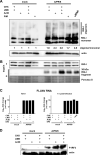
Fig. 1. Activation of RIG-I signaling by incoming influenza virus nucleocapsids
(A and B) RIG-I activity assays. A549 cells were pre-incubated for 1 h with inhibitors, inoculated with strain A/PR/8/34 (MOI 1) or left uninfected (mock) for 1 h at 4°C, incubated 1 h at 37°C, and analyzed. (A) Oligomerization assay. Lysates of cells treated with CHX (50 μg/ml), LMB (16 nM), ActD (1 μg/ml), or IVM (50 μM) were separated by native PAGE and immunostained for RIG-I. Actin served as loading control. (B) Conformational switch. Lysates as in (A) were subjected to limited trypsin digest and analyzed by RIG-I immunoblot. The Ponceau S protein stain (representative section shown) serves as loading control for the digested samples. (C) Quantification of total FLUAV segment 7 RNA by RT-qPCR. Input represents RNA amounts harvested after the 1 h inoculation period at 4°C. (D) IRF3 activation. A549 cells were pretreated with inhibitors, inoculated with strain A/PR/8/34 (MOI 1) or left uninfected (mock) for 1 h at 4°C, and incubated for 1 h under inhibitor treatment. Lysates from cells were separated by native PAGE and analyzed by immunoblotting for phosphorylated IRF3 (P-IRF3) and actin as described (Weber et al., 2013). See also Figures S1A-S1C.
Fig. 2
Fig. 2. RIG-I interacts with incoming influenza virus nucleocapsids and is activated in a 5’ppp-dsRNA-dependent manner
(A to C) CHX / LMB-treated A549 cells were infected with A/PR/8/34 (MOI 1) for 1 h. (A) Cells analyzed for RIG-I and FLUAV by 3D GSD superresolution immunofluorescence microscopy. Scale bar, 1 μm. Insets are digitally magnified and shown below the main image (taken from one individual cell). (B) Co-immunoprecipitation. Cell lysates were subjected to immunoprecipitation (IP) using antibodies against p21 (negative control), RIG-I, or FLUAV NP, and analyzed by immunoblot. Input control: 2% of the lysate. Asterisks (*) indicate unspecific bands. (C) Co-sedimentation assay. Cell lysates were separated by a discontinuous CsCl gradient (2% lysate as input control), and fractions analyzed by immunoblotting. (D) Conditions for activation of RIG-I by nucleocapsids in vitro. Dialyzed lysate of RIG-I-expressing S2 cells was mixed with nucleocapsids of strain A/PR/8/34 (RNPs) or a control preparation (CTRL) and supplemented with 1 mM ATP. The nucleocapsids had either been pretreated with 5 μg RNase A (A), 1 U RNase III (III), or 2 U Shrimp Alkaline Phosphatase (SAP), or left untreated (−), for 1 h at 37°C. RIG-I conformational switch was assayed after 1 h of nucleocapsid co-incubation at 37°C. See also Figures S2A-S2K.
Fig. 3
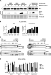
Fig. 3. Adaptive mutations in PB2 influence the activation of RIG-I by FLUAV nucleocapsids
(A) RIG-I activation by viruses with different PB2-627 signatures. Cells were infected with strains of A/quail/Shantou/2061/00 (H9N2), A/Thai/KAN-1/04 (H5N1), A/Hamburg/05/2009 (pH1N1), or A/WSN/33 (H1N1) containing avian-signature E or mammalian-signature K at PB2-627. Infections, CHX/LMB treatment and RIG-I conformational switch testing were performed as described for 1 and 2. (B) Quantification of virus RNAs by RT-qPCR for genomic segment 7. Input represents RNA amounts harvested after the 1-h infection period. (C) Co-sedimentation assay. Lysates of cells infected with PB2 variants of A/WSN/33 (H1N1) using our standard 1 h-protocol were separated by a CsCl gradient and analyzed by immunoblotting. (D) RIG-I-dependent IFN induction by incoming nucleocapsids. A549 cells were transfected with the indicated siRNAs or a negative control siRNA (CTRL). A549 cells siRNA-depleted of RIG-I or MDA5 were pretreated with CHX and LMB, and infected with FLUAV strains (MOI 1) for 16 h. IFN-β mRNA levels were determined by real-time RTPCR. See also Figures S3A-S3H.
Fig. 4
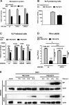
Fig. 4. RIG-I evasion by PB2-E627K
(A) Activity of A/WSN/33-based minireplicon systems containing PB2-627K, −627E, or no PB2 (−) in HEK293 wt, ΔRIG-I or ΔMDA5 cells. (B) Reporter activities in HEK293 cells producing VLPs containing nucleocapsids with the indicated PB2 signatures. (C) Reporter activities in wt and deletion cells infected with VLPs. Cells had been pretransfected with PB1, PA, NP, and matching PB2. (D) Multicycle virus kinetics. 293 wt or ΔRIG-I cells were infected with the indicated PB2 variants of strain A/Thai/KAN-1/04 (H5N1) at an MOI 0.0001. Virus yields were determined 24 h later by plaque assay. In all cases, mean and SDs from 3 independent experiments are shown. (E) Single-cycle virus kinetics. Cells were infected at an MOI of 1 and monitored for NP expression over time. See also Figures S4A-S4D.
Fig. 5
Fig. 5. Signaling-independent RIG-I effect on early infection
Single-cycle infection kinetics of A/WSN/33 on human cells lacking MAVS (A) or RIG-I (B), or on chicken DF-1 cells that naturally lack RIG-I (C). The ΔRIG-I and the DF-1 cells were transiently transfected with plasmids encoding GFP (negative control), FLAG-RIG-I wt, or FLAG-RIG-I K270A, as indicated. Overexpression was controlled using antibodies against GFP or the N-terminal Flag tag of the RIG-I constructs. See also Figure S5.
Fig. 6
Fig. 6. Effect of the PB2 627 signature on protein-protein interactions
(A) NP immunoprecipitation. Cells were CHX / LMB treated and infected with A/WSN/33 strains carrying PB2-627K or 627E as described for 1A. Lysates were immunoprecipitated 1 h later with anti-NP and immunoblotted as indicated. Normalized quantifications of the immunoprecipitated proteins are shown below. Note that amounts of viral input proteins are too low to be detected in the total extracts. (B) RIG-I immunoprecipitations from infected cells. HEK293 cells were infected with A/WSN/33 PB2 variants as described for 1A. Immunoprecipations with anti-RIG-I and immunoblotting were performed as indicated for 2B. (C) RIG-I immunoprecipitations of recombinant nucleocapsids. HEK293 cells were transfected with A/WSN/33 NP combined with GFP or the PB2 variants (left panel), or with all A/WSN/33 minireplicon plasmids (right panel). Immunoprecipations with anti-RIG-I were performed as indicated for (B). (D) Polymerase destabilization. A549 cells were infected with PB2 variants of strain A/WSN/33 (MOI 1), treated with peptides Borna-X-Tat (CTRL) or PB11-15 T6Y-Tat (PB1-T6Y), and tested for RIG-I conformational switch 1 h post-infection. See also Figures S6A-S6C.

Comment in

  • RIG-I works double duty.
    Chan YK, Gack MU. Chan YK, et al. Cell Host Microbe. 2015 Mar 11;17(3):285-287. doi: 10.1016/j.chom.2015.02.014. Cell Host Microbe. 2015. PMID: 25766287 Free PMC article.

Similar articles

Cited by

References

    1. Barber MR, Aldridge JR, Jr., Webster RG, Magor KE. Association of RIG-I with innate immunity of ducks to influenza. Proceedings of the National Academy of Sciences of the United States of America. 2010;107:5913–5918. - PMC - PubMed
    1. Baron J, Tarnow C, Mayoli-Nussle D, Schilling E, Meyer D, Hammami M, Schwalm F, Steinmetzer T, Guan Y, Garten W, et al. Matriptase, HAT, and TMPRSS2 activate the hemagglutinin of H9N2 influenza A viruses. Journal of virology. 2013;87:1811–1820. - PMC - PubMed
    1. Baum A, Sachidanandam R, Garcia-Sastre A. Preference of RIG-I for short viral RNA molecules in infected cells revealed by next-generation sequencing. Proceedings of the National Academy of Sciences of the United States of America. 2010;107:16303–16308. - PMC - PubMed
    1. Cauldwell AV, Long JS, Moncorge O, Barclay WS. Viral determinants of influenza A virus host range. The Journal of general virology. 2014;95:1193–1210. - PubMed
    1. Cauldwell AV, Moncorge O, Barclay WS. Unstable polymerase nucleoprotein interaction is not responsible for avian influenza virus polymerase restriction in human cells. Journal of virology. 2013;87:1278–1284. - PMC - PubMed

Publication types

MeSH terms