

Dynamic imaging of accommodation by swept-source anterior segment optical coherence tomography
- PMID: 25704218
- PMCID: PMC4699182
- DOI: 10.1016/j.jcrs.2014.09.034
Dynamic imaging of accommodation by swept-source anterior segment optical coherence tomography
Abstract
Purpose: To study the accommodation process in normal eyes using a commercially available clinical system based on swept-source anterior segment optical coherence tomography (AS-OCT).
Setting: Ophthalmology Department, University of Parma, Italy.
Design: Evaluation of diagnostic technology.
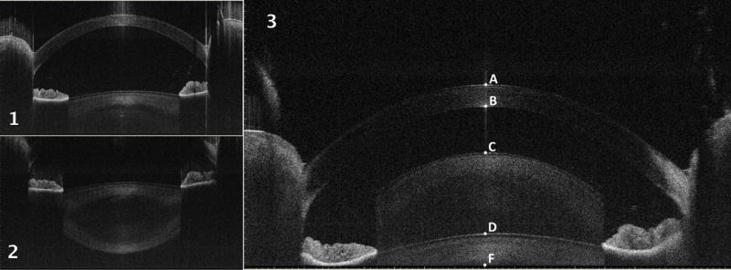
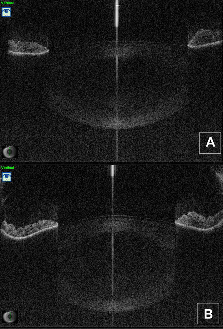
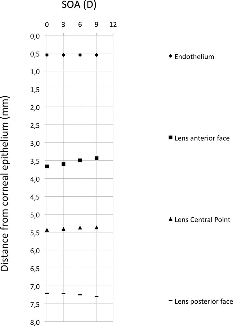
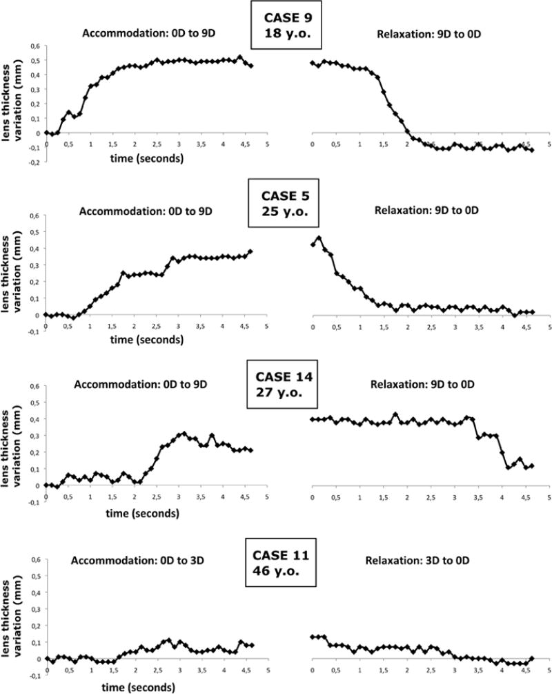
Methods: Right eyes were analyzed using swept-source AS-OCT (Casia SS-1000). The optical vergence of the internal coaxial fixation target was adjusted during imaging to obtain monocular accommodation stimuli with different amplitudes (0, 3.0, 6.0, and 9.0 diopters [D]). Overlapping of real and conjugate OCT images enabled imaging of all the anterior segment optical surfaces in a single frame. Central corneal thickness (CCT), anterior chamber depth (ACD), and lens thickness were extracted from the OCT scans acquired at different static accommodation stimulus amplitudes. The crystalline lens was analyzed dynamically during accommodation and disaccommodation by acquiring sequential OCT images of the anterior segment at a rate of 8 frames per second. The lens thickness was extracted from the temporal sequence of OCT images and plotted as a function of time.
Results: The study analyzed 14 eyes of 14 subjects aged 18 to 46 years. During accommodation, the decrease in the ACD was statistically significant (P < .05), as were the increase in the lens thickness (P < .001) and the slight movement forward of the lens central point (P < .01). The CCT and anterior chamber width measurements did not change statistically significantly during accommodation. The lens thickness at 0 D was positively correlated with age (P < .01).
Conclusion: High-resolution real-time imaging and biometry of the accommodating anterior segment can be effectively performed using a commercially available swept-source AS-OCT clinical device.
Financial disclosure: No author has a financial or proprietary interest in any material or method mentioned.
Copyright © 2015 ASCRS and ESCRS. Published by Elsevier Inc. All rights reserved.
Conflict of interest statement
Figures
Similar articles
-
Non-invasive measurements of the dynamic changes in the ciliary muscle, crystalline lens morphology, and anterior chamber during accommodation with a high-resolution OCT.Graefes Arch Clin Exp Ophthalmol. 2017 Jul;255(7):1385-1394. doi: 10.1007/s00417-017-3663-4. Epub 2017 Apr 20. Graefes Arch Clin Exp Ophthalmol. 2017. PMID: 28424868
-
Anterior segment biometry during accommodation imaged with ultralong scan depth optical coherence tomography.Ophthalmology. 2012 Dec;119(12):2479-85. doi: 10.1016/j.ophtha.2012.06.041. Epub 2012 Aug 17. Ophthalmology. 2012. PMID: 22902211 Free PMC article.
-
Static and dynamic analysis of the anterior segment with optical coherence tomography.J Cataract Refract Surg. 2004 Sep;30(9):1843-50. doi: 10.1016/j.jcrs.2004.05.024. J Cataract Refract Surg. 2004. PMID: 15342045
-
Value of optical coherence tomography for anterior segment surgery.J Cataract Refract Surg. 2010 Jul;36(7):1213-29. doi: 10.1016/j.jcrs.2010.05.002. J Cataract Refract Surg. 2010. PMID: 20610103 Review.
-
Clinical utility of anterior segment swept-source optical coherence tomography in glaucoma.Oman J Ophthalmol. 2016 Jan-Apr;9(1):3-10. doi: 10.4103/0974-620X.176093. Oman J Ophthalmol. 2016. PMID: 27013821 Free PMC article. Review.
Cited by
-
Effect of age in the ciliary muscle during accommodation: Sectorial analysis.J Optom. 2019 Jan-Mar;12(1):14-21. doi: 10.1016/j.optom.2018.01.001. Epub 2018 Apr 4. J Optom. 2019. PMID: 29627301 Free PMC article.
-
Non-invasive measurements of the dynamic changes in the ciliary muscle, crystalline lens morphology, and anterior chamber during accommodation with a high-resolution OCT.Graefes Arch Clin Exp Ophthalmol. 2017 Jul;255(7):1385-1394. doi: 10.1007/s00417-017-3663-4. Epub 2017 Apr 20. Graefes Arch Clin Exp Ophthalmol. 2017. PMID: 28424868
-
Repeatability and agreement of two anterior segment OCT in myopic patients before implantable collamer lenses implantation.Int J Ophthalmol. 2020 Apr 18;13(4):625-631. doi: 10.18240/ijo.2020.04.15. eCollection 2020. Int J Ophthalmol. 2020. PMID: 32399415 Free PMC article.
-
Dynamic refraction and anterior segment OCT biometry during accommodation.Biomed Opt Express. 2024 Apr 5;15(5):2876-2889. doi: 10.1364/BOE.512193. eCollection 2024 May 1. Biomed Opt Express. 2024. PMID: 38855690 Free PMC article.
-
Comparison of anterior segment and lens biometric measurements in patients with cataract.Graefes Arch Clin Exp Ophthalmol. 2020 Jan;258(1):137-146. doi: 10.1007/s00417-019-04482-0. Epub 2019 Oct 20. Graefes Arch Clin Exp Ophthalmol. 2020. PMID: 31631237
References
-
- Helmholtz H. Ueber die Accommodation des Auges. Albrecht von Graefes Arch Ophthalmol. 1855;2:1–74.
-
- Charman WN. The eye in focus: accommodation and presbyopia. Clinical & experimental optometry: journal of the Australian Optometrical Association. 2008;91:207–225. - PubMed
-
- Sheppard AL, Evans CJ, Singh KD, Wolffsohn JS, Dunne MC, Davies LN. Three-dimensional magnetic resonance imaging of the phakic crystalline lens during accommodation. Invest Ophthalmol Vis Sci. 2011;52:3689–3697. - PubMed
-
- Kasthurirangan S, Markwell EL, Atchison DA, Pope JM. MRI study of the changes in crystalline lens shape with accommodation and aging in humans. J Vis. 2011;11:1–16. - PubMed
-
- Hermans EA, Pouwels PJ, Dubbelman M, Kuijer JP, van der Heijde RG, Heethaar RM. Constant volume of the human lens and decrease in surface area of the capsular bag during accommodation: an MRI and Scheimpflug study. Invest Ophthalmol Vis Sci. 2009;50:281–289. - PubMed
Publication types
MeSH terms
Grants and funding
LinkOut - more resources
Full Text Sources
Other Literature Sources
Research Materials
