. 2015 Feb 23:6:6333.
doi: 10.1038/ncomms7333.

Cyclic stretching of soft substrates induces spreading and growth

Affiliations

Cyclic stretching of soft substrates induces spreading and growth

Yidan Cui et al. Nat Commun. .

Abstract

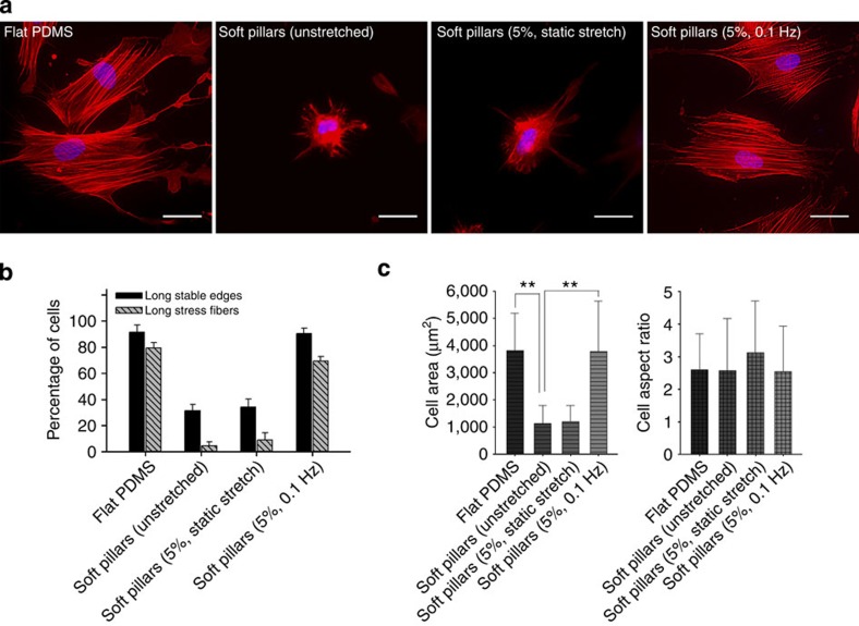
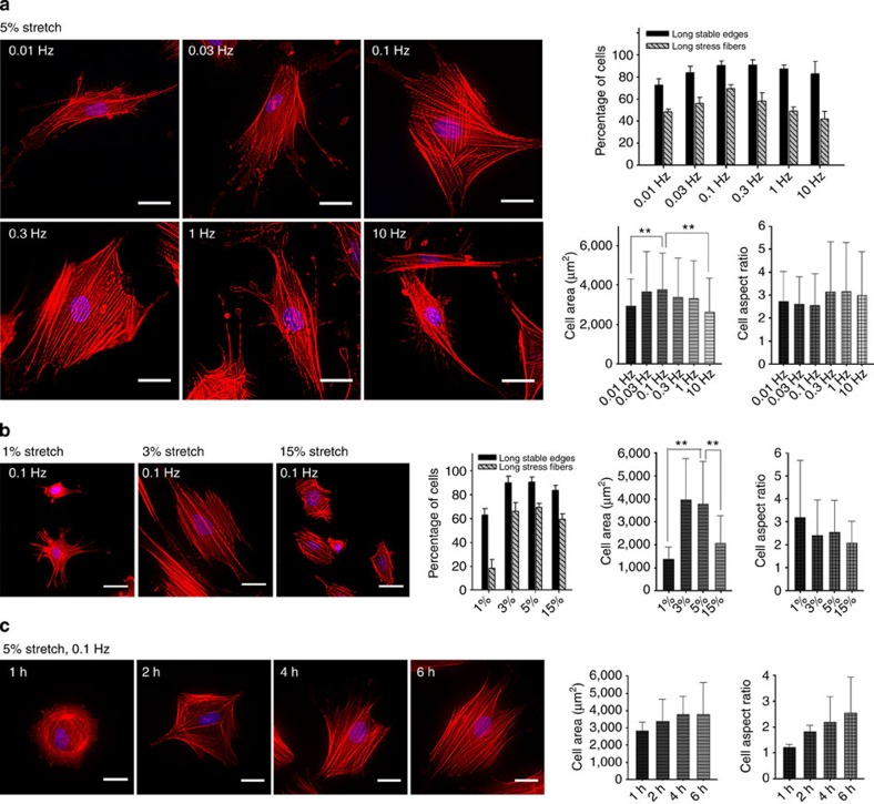
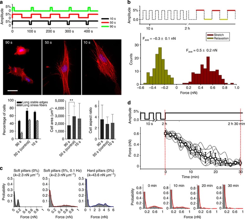
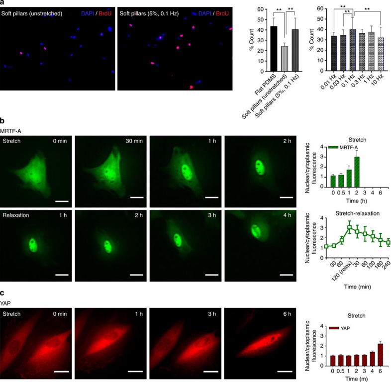
In the body, soft tissues often undergo cycles of stretching and relaxation that may affect cell behaviour without changing matrix rigidity. To determine whether transient forces can substitute for a rigid matrix, we stretched soft pillar arrays. Surprisingly, 1-5% cyclic stretching over a frequency range of 0.01-10 Hz caused spreading and stress fibre formation (optimum 0.1 Hz) that persisted after 4 h of stretching. Similarly, stretching increased cell growth rates on soft pillars comparative to rigid substrates. Of possible factors linked to fibroblast growth, MRTF-A (myocardin-related transcription factor-A) moved to the nucleus in 2 h of cyclic stretching and reversed on cessation; but YAP (Yes-associated protein) moved much later. Knockdown of either MRTF-A or YAP blocked stretch-dependent growth. Thus, we suggest that the repeated pulling from a soft matrix can substitute for a stiff matrix in stimulating spreading, stress fibre formation and growth.


Figures

Figure 1
Figure 1. Effect of substrate rigidity and stretching type on cell spreading and stress fibre formation.
(a) Representative images of PMEFs on flat PDMS (polydimethylsiloxane) and soft pillars made of PDMS with and without stretching. After being seeded onto each substrate, cells were incubated with and without stretching for 6 h before being stained with rhodamine-phalloidin (RP) and 4′, 6-diamidino-2-phenylindole (DAPI). Scale bar=30 μm. Flat PDMS served as a control. (b) Percentage of PMEFs with long stable edges and long stress fibres and (c) cell area and cell aspect ratio on each substrate: flat PDMS (n=153 cells); unstretched soft pillars (n=159); soft pillars with 5% static stretching (n=105); soft pillars with 5% cyclic stretching at 0.1 Hz (n=175). Error bars, s.e.m. **P<0.01; Student’s t-test. Experiments were repeated at least three times.
Figure 2
Figure 2. Effect of strain frequency and magnitude of cyclic stretching on cell spreading and stress fiber formation on soft pillars.
(a) PMEFs on soft pillars with 5% cyclic stretching for 6 h at different frequencies: 0.01 Hz (n=126 cells); 0.03 Hz (n=92); 0.1 Hz (n=175); 0.3 Hz (n=171); 1 Hz (n=134); 10 Hz (n=174). (b) Cyclic stretch (0.1 Hz) for 6 h at various amplitudes: 1% (n=104 cells); 3% (n=132 cells); 5% (n=175); 15% (n=175). (c) 5% Cyclic stretching (0.1 Hz) for various periods of time: 1 h (n=113 cells); 2 h (n=95); 4 h (n=101 cells); 6 h (n=175). Error bars; s.e.m. **P<0.01; Student’s t-test. Cells were stained with RP and DAPI after stretching. Scale bar=30 μm. Experiments were repeated at least three times.
Figure 3
Figure 3. Effects of relaxation time of cyclic stretching on cell morphology and contractile responses of cells to cyclic stretch.
(a) PMEFs on soft pillars with 5% cyclic stretching (0.01 Hz) for 6 h at different relaxation times: 10 s (n=95 cells); 50 s (n=126 cells); 90 s (n=114 cells). Cells were fixed 6 h after seeding and stained with RP and DAPI. Scale bar=30 μm. Error bars; s.e.m. **P<0.01; Student’s t-test. (b) Distributions of forces calculated from single stretch–relaxation event. Cells were first stretched for 2 h (5%, 0.1 Hz) after seeding and then displacements of pillars were calculated from single stretch–relaxation event (n=12 cells). Only the pillars near the cell edge were calculated. Fave means average forces measured on stretch–relaxation. (c) Distributions of forces on soft or hard pillars (n>300 pillars). (d) Changes in forces over time on changing from 5% cyclic stretching after 2 h to 5% static stretching (n>300 pillars). Experiments were repeated at least three times and showed similar results.
Figure 4
Figure 4. Effect of cyclic stretching on cell proliferation.
(a) Percentage of cells costained with 5-bromo-2′-deoxyuridine (BrdU) and DAPI on different stretching types and frequencies: unstretched flat PDMS (n=140 cells), unstretched soft pillars (n=142), 5% cyclic stretched soft pillars at 0.1 Hz (n=74), 0.01 Hz (n=93), 0.03 Hz (n=66), 0.3 Hz (n=106), 1 Hz (n=59) and 10 Hz (n=79). Error bars; s.e.m. **P<0.01; Student’s t-test. Time-dependent translocation of (b) GFP–MRTF-A (green fluorescent protein tagged Myocardin-related transcription factor-A) and (c) YAP–GFP (green fluorescent protein tagged Yes-associated protein) by cyclic stretch (5%, 0.1 Hz). Plasmids were transient transfected into PMEFs through electroporation at least 24 h before imaging. Stretching was applied 2 h after the seeding of cells in the chamber. Over 20 cells were analysed for each protein. Experiments were repeated at least three times and showed similar results. Scale bar=20 μm.

References

    1. Vogel V. & Sheetz M. Local force and geometry sensing regulate cell functions. Nat. Rev. Mol. Cell Biol. 7, 265–275 (2006). - PubMed
    1. Wang J. H. & Thampatty B. P. An introductory review of cell mechanobiology. Biomech. Model. Mechanobiol. 5, 1–16 (2006). - PubMed
    1. Georges P. C. & Janmey P. A. Cell type-specific response to growth on soft materials. J. Appl. Physiol. 98, 1547–1553 (2005). - PubMed
    1. Tamada M., Sheetz M. P. & Sawada Y. Activation of a signaling cascade by cytoskeleton stretch. Dev. Cell 7, 709–718 (2004). - PubMed
    1. Schrader J. et al.. Matrix stiffness modulates proliferation, chemotherapeutic response, and dormancy in hepatocellular carcinoma cells. Hepatology 53, 1192–1205 (2011). - PMC - PubMed

Publication types