

Actinomyces-induced inflammatory myofibroblastic tumor of the colon: A rare cause of an abdominal mass: Akbulut et al. inflammatory myofibroblastictumor due to actinomyces spp
- PMID: 25704558
- PMCID: PMC4392327
- DOI: 10.1016/j.ijscr.2015.02.014
Actinomyces-induced inflammatory myofibroblastic tumor of the colon: A rare cause of an abdominal mass: Akbulut et al. inflammatory myofibroblastictumor due to actinomyces spp
Abstract
Introduction: Inflammatory myofibroblastic tumors (IMFTs) are neoplastic lesions that are either benign or have low-grade malignancy potential. Although the etiopathogenesis is not entirely clear, many factors play a role in their development, including trauma, autoimmune disorders, and infectious and inflammatory processes. However, IMFTs caused by Actinomyces spp. infection are rare, with a limited number of cases reported in the literature.
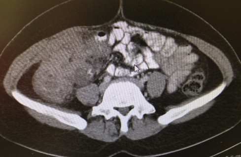
Presentation of case: A 30-year-old woman was admitted to our clinic with abdominal pain and a palpable abdominal mass. Contrast-enhanced computed tomography revealed a tumoral lesion (11×10×7cm) in the right colon. A right hemicolectomy and ileocolic anastomosis were performed, during which almost complete obstruction of the lumen by the 7.5×7.0×5.0cm tumor was observed. Histopathology and immunohistochemical findings revealed that the tumor was consistent with an IMFT that developed from an Actinomyces infection. The patient was then placed on amoxicillin and doxycycline therapy.
Conclusion: This case demonstrates that the development of IMFT secondary to actinomycosis is difficult to predict in the preoperative period. Once an exact diagnosis is confirmed by histopathologic examination, affected patients should receive prolonged antibiotherapy.
Keywords: Actinomycosis; Colon; Immunhistochemical stain; Inflammatory myofibroblastic tumor.
Copyright © 2015 The Authors. Published by Elsevier Ltd.. All rights reserved.
Figures


References
-
- Yagmur Y., Akbulut S., Gumus S. Mesenteric inflammatory pseudotumor: a case report and comprehensive literature review. J. Gastrointest. Cancer. 2014;45(4):414–420. - PubMed
-
- Das N., Lee J., Madden M., Elliot C.S., Bateson P., Gilliland R. A rare case of abdominal actinomycosis presenting as an inflammatory pseudotumour. Int. J. Colorectal. Dis. 2006;21(5):483–484. - PubMed
-
- Evans J., Chan C., Gluch L., Fielding I., Eckstein R. Inflammatory pseudotumour secondary to actinomyces infection. Aust. N. Z. J. Surg. 1999;6:467–469. - PubMed
-
- Radhi J., Hadjis N., Anderson L., Burbridge B., Ali K. Retroperitoneal actinomycosis masquerading as inflammatory pseudotumor. J. Pediatr. Surg. 1997;32(4):618–620. - PubMed
LinkOut - more resources
Full Text Sources
Other Literature Sources
Research Materials
Miscellaneous
