Review
. 2016 Feb;89(2):111-22.
doi: 10.1002/cyto.a.22647. Epub 2015 Feb 20.

Microparticle analysis in disorders of hemostasis and thrombosis

Affiliations
Review

Microparticle analysis in disorders of hemostasis and thrombosis

Micah J Mooberry et al. Cytometry A. 2016 Feb.

Abstract

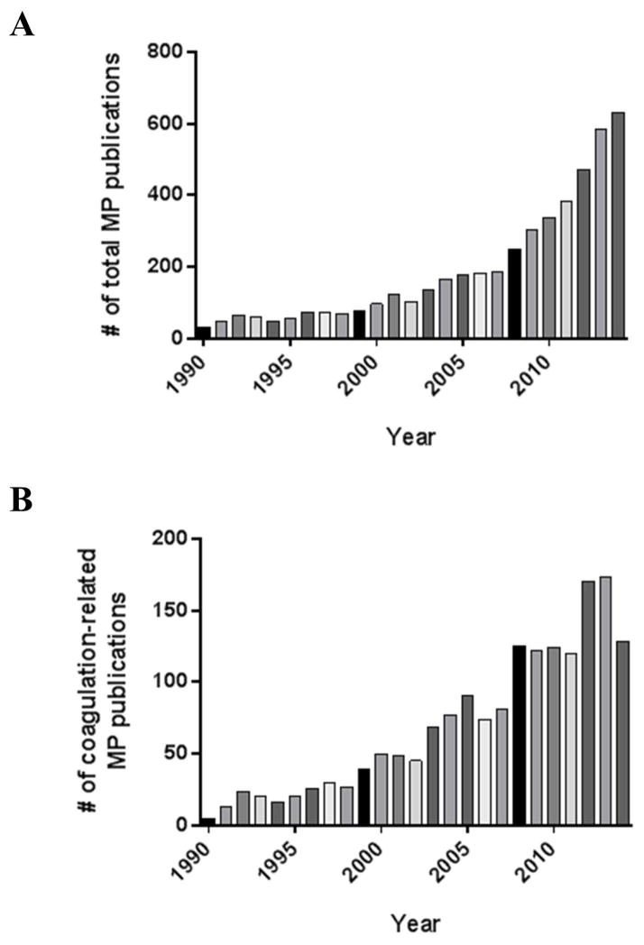
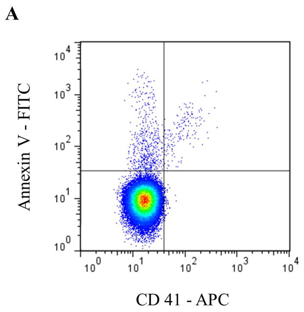
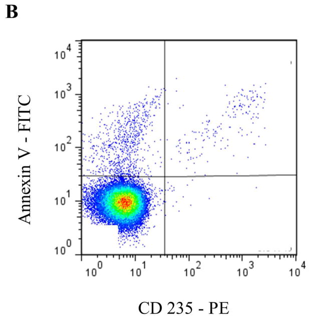
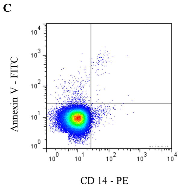
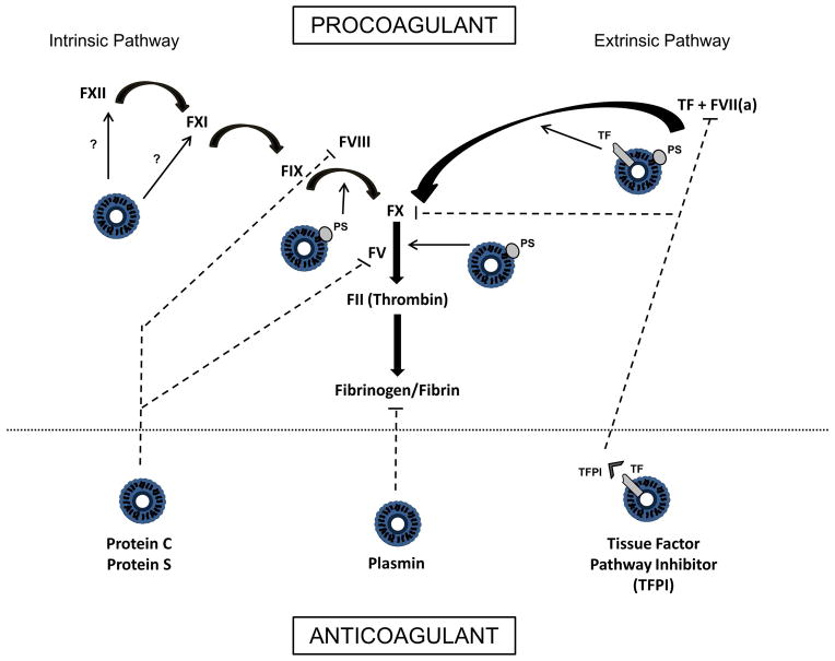
Microparticles (MPs) are submicron vesicles released from the plasma membrane of eukaryotic cells in response to activation or apoptosis. MPs are known to be involved in numerous biologic processes, including inflammation, the immune response, cancer metastasis, and angiogenesis. Their earliest recognized and most widely accepted role, however, is the ability to promote and support the process of blood coagulation. Consequently, there is ongoing interest in studying MPs in disorders of hemostasis and thrombosis. Both phosphatidylserine (PS) exposure and the presence of tissue factor (TF) in the MP membrane may account for their procoagulant properties, and elevated numbers of MPs in plasma have been reported in numerous prothrombotic conditions. To date, however, there are few data on true causality linking MPs to the genesis of thrombosis. A variety of methodologies have been employed to characterize and quantify MPs, although detection is challenging due to their submicron size. Flow cytometry (FCM) remains the most frequently utilized strategy for MP detection; however, it is associated with significant technological limitations. Additionally, preanalytical and analytical variables can influence the detection of MPs by FCM, rendering data interpretation difficult. Lack of methodologic standardization in MP analysis by FCM confounds the issue further, although efforts are currently underway to address this limitation. Moving forward, it will be important to address these technical challenges as a scientific community if we are to better understand the role that MPs play in disorders of hemostasis and thrombosis.

Keywords: coagulation; flow cytometry; hemostasis; microparticles; microvesicles; thrombosis.


Conflict of interest statement

The authors have no conflicts of interest to disclose.

Figures

Figure 1
Figure 1
Number of (A) total MP publications, and (B) specific coagulation-related MP publications by year since 1990. Total MP publication numbers were acquired utilizing a PubMed search for articles from 1990–2014 with keywords “microparticles or microvesicles”, while excluding studies related to pharmacology, drug delivery and non-biological entities. Coagulation-related MP publication numbers were acquired utilizing a PubMed search for articles from 1990–2014 with keywords “microparticles or microvesicles” in conjunction with coagulation specific terms such as “thrombosis” and “hemostasis”.
Figure 2
Figure 2
Representative staining and gating strategies for A) platelet microparticles (PMP), B) red blood cell microparticles (RMP), and C) monocyte microparticles (MMP) analyzed in platelet free plasma on a Stratedigm S1000Ex flow cytometer. Fluorescent gating was performed within the MP size gate of 200–900 μm, which was initially set utilizing polystyrene beads (data not shown). PMP = dual positive Annexin V/CD41 events. RMP = dual positive Annexin V/CD235 events. MMP = dual positive Annexin V/CD14 events.
Figure 2
Figure 2
Representative staining and gating strategies for A) platelet microparticles (PMP), B) red blood cell microparticles (RMP), and C) monocyte microparticles (MMP) analyzed in platelet free plasma on a Stratedigm S1000Ex flow cytometer. Fluorescent gating was performed within the MP size gate of 200–900 μm, which was initially set utilizing polystyrene beads (data not shown). PMP = dual positive Annexin V/CD41 events. RMP = dual positive Annexin V/CD235 events. MMP = dual positive Annexin V/CD14 events.
Figure 3
Figure 3. Multifaceted role of MPs in coagulation processes
Simplified schemata of the coagulation cascade showing the different potential contributions of MPs. MPs support coagulation through exposure of phosphatidylserine (PS), which provides a catalytic surface for assembly of the coagulation complexes. Tissue factor (TF) bearing MPs can activate coagulation through the extrinsic pathway. MPs may also support coagulation through the intrinsic pathway, although the mechanism by which this occurs is not fully known. Anticoagulant properties of MPs include the ability to support Protein C/Protein S mediated regulation of coagulation, as well as tissue factor pathway inhibitor (TFPI) mediated inhibition of TF/VIIa activity and FX. MPs can also support plasmin generation, an enzyme that solubilizes and degrades clots. (Bolded arrows indicate activation steps [ie FXII activates FXI]. Dashed lines indicate inhibitory effects. Unbolded arrows emanating from MPs indicate areas of MP participation in coagulation activation processes.)

Similar articles

Cited by

References

    1. Chargaff E, West R. The biological significance of the thromboplastic protein of blood. J Biol Chem. 1946;166:189–97. - PubMed
    1. Wolf P. The nature and significance of platelet products in human plasma. Br J Haematol. 1967;13:269–88. - PubMed
    1. Tesse A, Martínez MC, Meziani F, Hugel B, Panaro MA, Mitolo V, Freyssinet JM, Andriantsitohaina R. Origin and Biological Significance of Shed-Membrane Microparticles. Endocrine, Metabolic & Immune Disorders - Drug Targets. 2006;6:287–94. - PubMed
    1. Zwaal RFA, Comfurius P, Bevers EM. Surface exposure of phosphatidylserine in pathological cells. Cell Mol Life Sci. 2005;62:971–88. - PMC - PubMed
    1. Thaler J, Ay C, Pabinger I. Clinical significance of circulating microparticles for venous thromboembolism in cancer patients. Hamostaseologie. 2012;32:127–31. - PubMed

Publication types

LinkOut - more resources