

The Molecular Mechanisms of Neural Flow Coupling: A New Concept
- PMID: 25704766
- PMCID: PMC5023998
- DOI: 10.1111/jon.12219
The Molecular Mechanisms of Neural Flow Coupling: A New Concept
Abstract
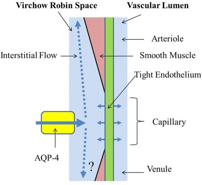
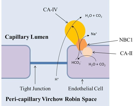
The phenomenon known as neural flow coupling (NFC) occurs at the capillary level where there are no known pressure controlling structures. Recent developments in advanced magnetic resonance imaging technologies have made possible in vivo direct investigations of water physiology that have shed new insight on the water dynamics of the cortical pericapillary space and their complex functionality in relation to NFC. Neural activities initiate a chain of events that ultimately affect NFC. First, neural activities generate extracellular acidification. Extracellular acidosis in turn produces inhibition of aquaporin-4 (AQP-4) located at the end feet of pericapillary astrocytes, the water channel which regulates water influx into the pericapillary space and, hence, interstitial flow. Reduction of pericapillary water pressure results in a negative balance between pericapillary and intraluminal capillary pressure, allowing for capillary caliber expansion. Proton permeability through the tight junctions of the blood brain barrier is significantly high owing to the Grotthuss proton "tunneling" mechanism and, therefore, carbonic anhydrase (CA) type IV (CA-IV) anchored to the luminal surface of brain capillaries functions as scavenger of extracellular protons. CA-IV inhibition by acetazolamide or carbon dioxide results in the accumulation of extracellular protons, causing AQP-4 inhibition and a secondary increase in rCBF.
Keywords: aquaporin 4; interstitial flow; neural activation; neuro flow coupling; protons.
© 2015 The Authors. Journal of Neuroimaging published by Wiley Periodicals, Inc. on behalf of American Society of Neuroimaging.
Figures


Similar articles
-
Aquaporin-4 Functionality and Virchow-Robin Space Water Dynamics: Physiological Model for Neurovascular Coupling and Glymphatic Flow.Int J Mol Sci. 2017 Aug 18;18(8):1798. doi: 10.3390/ijms18081798. Int J Mol Sci. 2017. PMID: 28820467 Free PMC article. Review.
-
Virchow-Robin space and aquaporin-4: new insights on an old friend.Croat Med J. 2014 Aug 28;55(4):328-36. doi: 10.3325/cmj.2014.55.328. Croat Med J. 2014. PMID: 25165047 Free PMC article. Review.
-
Aquaporin-4 facilitator TGN-073 promotes interstitial fluid circulation within the blood-brain barrier: [17O]H2O JJVCPE MRI study.Neuroreport. 2018 Jun 13;29(9):697-703. doi: 10.1097/WNR.0000000000000990. Neuroreport. 2018. PMID: 29481527 Free PMC article.
-
Water influx into cerebrospinal fluid is primarily controlled by aquaporin-4, not by aquaporin-1: 17O JJVCPE MRI study in knockout mice.Neuroreport. 2014 Jan 8;25(1):39-43. doi: 10.1097/WNR.0000000000000042. Neuroreport. 2014. PMID: 24231830 Free PMC article.
-
Fluid Dynamics Inside the Brain Barrier: Current Concept of Interstitial Flow, Glymphatic Flow, and Cerebrospinal Fluid Circulation in the Brain.Neuroscientist. 2019 Apr;25(2):155-166. doi: 10.1177/1073858418775027. Epub 2018 May 25. Neuroscientist. 2019. PMID: 29799313 Free PMC article. Review.
Cited by
-
Cell Settling, Migration, and Stochastic Cancer Gene Expression Suggest Potassium Membrane Flux May Initiate pH Reversal.Biomolecules. 2025 Aug 16;15(8):1177. doi: 10.3390/biom15081177. Biomolecules. 2025. PMID: 40867621 Free PMC article.
-
Role of Glutamatergic Excitotoxicity in Neuromyelitis Optica Spectrum Disorders.Front Cell Neurosci. 2019 Apr 12;13:142. doi: 10.3389/fncel.2019.00142. eCollection 2019. Front Cell Neurosci. 2019. PMID: 31031597 Free PMC article. Review.
-
Aquaporin-4 Functionality and Virchow-Robin Space Water Dynamics: Physiological Model for Neurovascular Coupling and Glymphatic Flow.Int J Mol Sci. 2017 Aug 18;18(8):1798. doi: 10.3390/ijms18081798. Int J Mol Sci. 2017. PMID: 28820467 Free PMC article. Review.
-
A Surgical Method to Improve the Homeostasis of CSF for the Treatment of Alzheimer's Disease.Front Aging Neurosci. 2016 Nov 2;8:261. doi: 10.3389/fnagi.2016.00261. eCollection 2016. Front Aging Neurosci. 2016. PMID: 27853433 Free PMC article.
-
Neurovascular Unit as a Source of Ischemic Stroke Biomarkers-Limitations of Experimental Studies and Perspectives for Clinical Application.Transl Stroke Res. 2020 Aug;11(4):553-579. doi: 10.1007/s12975-019-00744-5. Epub 2019 Nov 7. Transl Stroke Res. 2020. PMID: 31701356 Free PMC article. Review.
References
-
- Orešković D, Klarica M. The formation of cerebrospinal fluid: nearly a hundred years of interpretations and misinterpretations. Brain Res Rev 2010;64:241‐62. - PubMed
-
- Esiri MM, Gay D. Immunological and neuropathological significance of the Virchow‐Robin space. J Neurol Sci 1990;100:3‐8. - PubMed
-
- Weller RO. Pathology of cerebrospinal fluid and interstitial fluid of the CNS: significance for Alzheimer disease, prion disorders and multiple sclerosis. J Neuropathol Exp Neurol 1998;57:885‐94. - PubMed
Publication types
MeSH terms
Substances
LinkOut - more resources
Full Text Sources
Other Literature Sources
