. 2015:2015:464161.
doi: 10.1155/2015/464161. Epub 2015 Jan 29.

Millimeter Wave Treatment Inhibits Apoptosis of Chondrocytes via Regulation Dynamic Equilibrium of Intracellular Free Ca (2+)

Affiliations

Millimeter Wave Treatment Inhibits Apoptosis of Chondrocytes via Regulation Dynamic Equilibrium of Intracellular Free Ca (2+)

Jinxia Ye et al. Evid Based Complement Alternat Med. 2015.

Abstract

The molecular mechanisms of TNF-α-induced apoptosis of chondrocyte and the role of Ca(2+) mediating the effects of MW on TNF-α-induced apoptosis of chondrocytes remained unclear. In this study, we investigated the molecular mechanism underlying inhibiting TNF-α-induced chondrocytes apoptosis of MW. MTT assay, DAPI, and flow cytometry demonstrated that MW significantly increased cell activity and inhibited chromatin condensation accompanying the loss of plasma membrane asymmetry and the collapse of mitochondrial membrane potential. Our results also indicated that MW reduced the elevation of [Ca(2+)] i in chondrocytes by LSCM. Moreover, MW suppressed the protein levels of calpain, Bax, cytochrome c, and caspase-3, while the expressions of Bcl-2, collagen II, and aggrecan were increased. Our evidences indicated that MW treatment inhibited the apoptosis of chondrocytes through depression of [Ca(2+)] i . It also inhibited calpain activation, which mediated Bax cleavage and cytochrome c release and initiated the apoptotic execution phase. In addition, MW treatment increased the expression of collagen II and aggrecan of chondrocytes.


Figures

Figure 1
Figure 1
Identification of third generation of chondrocytes (×200). (a) Chondrocytes were stained using toluidine blue. Immunohistochemical staining of type II collagen demonstrated that (b) the cytoplasm of the negative group of chondrocytes was unstained, whereas (c) the cytoplasm of chondrocytes was stained.
Figure 2
Figure 2
Effect of millimeter wave on the morphological changes of chondrocytes (×200). (a) Untreated control chondrocytes; (b) chondrocytes treated with 10 ng/mL TNF-α for 8 h. (c) Chondrocytes treated with 10 ng/mL TNF-α for 8 h and then treated with millimeter wave for 30 min. The morphological changes of chondrocytes were observed using phase contrast microscopy.
Figure 3
Figure 3
Effect of TNF-α on chondrocyte viability. Chondrocytes were treated with 10 ng/mL TNF-α or TNF-α and millimeter wave (MW) for 4 hours. Cell viability was determined by the MTT assay. Data are averages with SD (error bars) from three independent experiments. P < 0.05, compared with the untreated control. P < 0.05, compared with the 10 ng/mL TNF-α group.
Figure 4
Figure 4
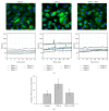
Effects of MW on intracellular [Ca2+]i expression in chondrocytes. (a) Fluorescence of intracellular [Ca2+]i was visualized by staining the chondrocytes with fluo-3/AM (green) and then counterstaining with Hoechst 33258 (blue). Images were captured under a confocal fluorescence microscope with a magnification of 400x. Five cells for each visual field were observed and the quantitative changes rule curve of intracellular [Ca2+]i fluorescence, which changed over time, was analyzed. (b) The changes in intensity of fluorescence of intracellular [Ca2+]i. Colorful curve representing the intracellular calcium concentration corresponds to the cell circled by line with same color. Data are averages with SD (error bars) from three independent experiments. * P < 0.05, compared with the untreated control. # P < 0.05, compared with the 10 ng/mL TNF-α group.
Figure 5
Figure 5
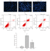
Effect of MW on cell apoptosis in chondrocytes. Chondrocytes were treated with 10 ng/mL TNF-α or TNF-α and MW, collected and stained with Hoechst 33258, and observed under a fluorescence microscope using Annexin-V+/PI and flow cytometry (FCM) analysis. (a) Visible condensed nuclei of chondrocytes stained with Hoechst 33258. (b) Apoptosis of chondrocytes analyzed by FACS. Images are representative of three independent experiments. (c) Quantification of FACS analysis. Data are averages with SD (error bars) from three independent experiments. * P < 0.05, compared with the untreated control. # P < 0.05, compared with the 10 ng/mL TNF-α group.
Figure 6
Figure 6
Effect of MW on the loss of mitochondrial membrane potential in chondrocytes. Chondrocytes were treated with 10 ng/mL TNF-α or TNF-α and MW and stained with JC-1. (a) The mean JC-1 fluorescence intensity was detected using FACS analysis. Data shown are representative of three independent experiments. (b) Quantification of FACS analysis. Data are averages with SD (error bars) from three independent experiments. * P < 0.05, compared with the untreated control. # P < 0.05, compared with the 10 ng/mL TNF-α group.
Figure 7
Figure 7
Effect of MW on the expression of calpain, Bax, Bcl-2, cytochrome c, caspase-3, collagen II, and aggrecan in chondrocytes. (a) The protein expression levels of calpain, Bax, Bcl-2, cytochrome c, caspase-3, collagen II, and aggrecan were analyzed by Western blotting. β-actin was used as the internal control. (b) Semiquantification of protein blots shown in (a). Data are averages with SD (error bars) from three independent experiments. * P < 0.05; ** P < 0.01, compared with the untreated control. # P < 0.05, ## P < 0.01, compared with the 10 ng/mL TNF-α group.

Similar articles

Cited by

References

    1. Goldring M. B., Goldring S. R. Articular cartilage and subchondral bone in the pathogenesis of osteoarthritis. Annals of the New York Academy of Sciences. 2010;1192(1):230–237. doi: 10.1111/j.1749-6632.2009.05240.x. - DOI - PubMed
    1. Zintzaras E., Kitsios G. D., Ziogas D. C., Rodopoulou P., Karachalios T. Field synopsis and synthesis of genetic association studies in osteoarthritis: the CUMAGAS-OSTEO information system. The American Journal of Epidemiology. 2010;171(8):851–858. doi: 10.1093/aje/kwq016. - DOI - PubMed
    1. Calich A. L. G., Domiciano D. S., Fuller R. Osteoarthritis: can anti-cytokine therapy play a role in treatment? Clinical Rheumatology. 2010;29(5):451–455. doi: 10.1007/s10067-009-1352-3. - DOI - PubMed
    1. Schulze-Tanzil G. Activation and dedifferentiation of chondrocytes: implications in cartilage injury and repair. Annals of Anatomy. 2009;191(4):325–338. doi: 10.1016/j.aanat.2009.05.003. - DOI - PubMed
    1. Ferraccioli G., Gremese E. Thrombogenicity of TNFα in rheumatoid arthritis defined through biological probes: TNFα blockers. Autoimmunity Reviews. 2004;3(4):261–266. doi: 10.1016/j.autrev.2003.09.004. - DOI - PubMed

LinkOut - more resources