Review
. 2014 Dec 10:3:27.
doi: 10.1186/2047-9158-3-27. eCollection 2014.

Aldehyde Dehydrogenase 1 making molecular inroads into the differential vulnerability of nigrostriatal dopaminergic neuron subtypes in Parkinson's disease

Affiliations
Review

Aldehyde Dehydrogenase 1 making molecular inroads into the differential vulnerability of nigrostriatal dopaminergic neuron subtypes in Parkinson's disease

Huaibin Cai et al. Transl Neurodegener. .

Abstract

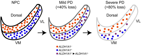
A preferential dysfunction/loss of dopaminergic (DA) neurons in the substantia nigra pars compacta (SNpc) accounts for the main motor symptoms of Parkinson's disease (PD), the most common degenerative movement disorder. However, the neuronal loss is not stochastic, but rather displays regionally selectivity, indicating the existence of different DA subpopulations in the SNpc. To identify the underlying molecular determinants is thereby instrumental in understanding the pathophysiological mechanisms of PD-related neuron dysfunction/loss and offering new therapeutic targets. Recently, we have demonstrated that aldehyde dehydrogenase 1 (ALDH1A1) is one such molecular determinant that defines and protects an SNpc DA neuron subpopulation preferentially affected in PD. In this review, we provide further analysis and discussion on the roles of ALDH1A1 in the function and survival of SNpc DA neurons in both rodent and human brains. We also explore the feasibility of ALDH1A1 as a potential biomarker and therapeutic target for PD.

Keywords: Aging; Aldehyde dehydrogenase 1; Dopaminergic neuron; Neurodegeneration; Parkinson’s disease; Substantia nigra pars compacta; α-synuclein.


Figures

Figure 1
Figure 1
Selective loss of ALDH1A1-positive subtype nigrostriatal DA neurons in PD. Cartoons illustrate the distribution of ALDH1A1–negative (ALDH1A1, red) and –positive (ALDH1A1+, blue) DA neurons, as well as ALDH1A1+ neurons that lose ALDH1A1 expression (ALDH1A1+➔ ALDH1A1, red with blue outline) in the SNpc of non-pathology control (NPC) and PD brains with mild and severe depigmentation. VL: ventral lateral, VM: ventral medial.
Figure 2
Figure 2
Expression of Aldh and Akr family genes in the mouse SNpc DA neurons. (A, B) RNA sequencing reveals the expression of Aldh (A) and Akt (B) family genes in SNpc DA neurons of 12-month-old wild type mice. Two independent sets of SNpc RNA samples were analyzed.
Figure 3
Figure 3
Dopamine metabolism and selective loss of SNpc DA neurons. Cartoon proposes the accumulation of cytosolic DOPAL triggers the degeneration of SNpc DA neurons in PD. Tyrosine hydroxylase (TH) and aromatic L-amino acid decarboxylase (AADC) mediate the synthesis of dopamine from tyrosine (Tyr) in the cytosol. Cytosolic dopamine is then immediately sequestered into the synaptic vesicles (SVs) by dopamine transporter VMAT2 for release. DAT mediates the reuptake of dopamine from extracellular space into the DA terminals. Leakage of dopamine from SVs also contributes to the cytosolic dopamine levels. MAO and ALDH1A1 are main enzymes for the degradation of cytosolic dopamine in DA neurons. A lack of ALDH1A1 may lead to a cytotoxic build-up of DOPAL which triggers the reactive oxygen species (ROS) production, protein adducts and α-synuclein aggregation, and eventually leads to cell death.
Figure 4
Figure 4
ALDH1A7 is highly homologous to ALDH1A1 in the mouse genome. (A) Diagrams depict the genomic structures of human ALDH1A1 (hALDH1A1) and mouse Aldh1a1 (mAldh1a1) and Aldh1a7 (mAldh1a7). Arrows point to the direction of transcription. (B) Table shows the percentage of identical amino acids shared between hALDH1A1, mALDH1A1, and mALDH1A7 proteins. (C) Western blot shows the residual proteins recognized by an ALDH1A1 antibody in both the midbrain (MB) and striatum of Aldh1a1 –/– mice.

References

    1. Parkinson J. An Essay On The Shaking Palsy. London: Sherwood, Nelly and Jones; 1817.
    1. Lewis PD. Parkinsonism–neuropathology. Br Med J. 1971;3(5776):690–692. doi: 10.1136/bmj.3.5776.690. - DOI - PMC - PubMed
    1. Cotzias GC. L-Dopa for Parkinsonism. N Engl J Med. 1968;278(11):630. - PubMed
    1. Pankratz ND, Wojcieszek J, Foroud T. Parkinson Disease Overview. In: Pagon RA, Adam MP, Bird TD, Dolan CR, Fong CT, Stephens K, editors. GeneReviews. Seattle: University of Washington; 1993. pp. 1993–2014. - PubMed
    1. Mercuri NB, Bernardi G. The 'magic' of L-dopa: why is it the gold standard Parkinson's disease therapy? Trends Pharmacol Sci. 2005;26(7):341–344. doi: 10.1016/j.tips.2005.05.002. - DOI - PubMed

LinkOut - more resources