A suspicious pancreatic mass in chronic pancreatitis: pancreatic actinomycosis
- PMID: 25705533
- PMCID: PMC4332457
- DOI: 10.1155/2015/767365
A suspicious pancreatic mass in chronic pancreatitis: pancreatic actinomycosis
Abstract
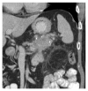
Introduction. Pancreatic actinomycosis is a chronic infection of the pancreas caused by the suppurative Gram-positive bacterium Actinomyces. It has mostly been described in patients following repeated main pancreatic duct stenting in the context of chronic pancreatitis or following pancreatic surgery. This type of pancreatitis is often erroneously interpreted as pancreatic malignancy due to the specific invasive characteristics of Actinomyces. Case. A 64-year-old male with a history of chronic pancreatitis and repeated main pancreatic duct stenting presented with weight loss, fever, night sweats, and abdominal pain. CT imaging revealed a mass in the pancreatic tail, invading the surrounding tissue and resulting in splenic vein thrombosis. Resectable pancreatic cancer was suspected, and pancreatic tail resection was performed. Postoperative findings revealed pancreatic actinomycosis instead of neoplasia. Conclusion. Pancreatic actinomycosis is a rare type of infectious pancreatitis that should be included in the differential diagnosis when a pancreatic mass is discovered in a patient with chronic pancreatitis and prior main pancreatic duct stenting. Our case emphasizes the importance of pursuing a histomorphological confirmation.
Figures


References
-
- Sahay S. J., Gonzalez H. D., Luong T. V., Rahman S. H. Pancreatic actinomycosis as a cause of retroperitoneal fibrosis in a patient with chronic pancreatitis. Case report and literature review. Journal of the Pancreas. 2010;11(5):477–479. - PubMed
LinkOut - more resources
Full Text Sources
Other Literature Sources

