. 2014;2(6):1-22.
doi: 10.1128/microbiolspec.MDNA3-0024-2014..

Retroviral Integrase Structure and DNA Recombination Mechanism

Affiliations

Retroviral Integrase Structure and DNA Recombination Mechanism

Alan Engelman et al. Microbiol Spectr. 2014.

Abstract

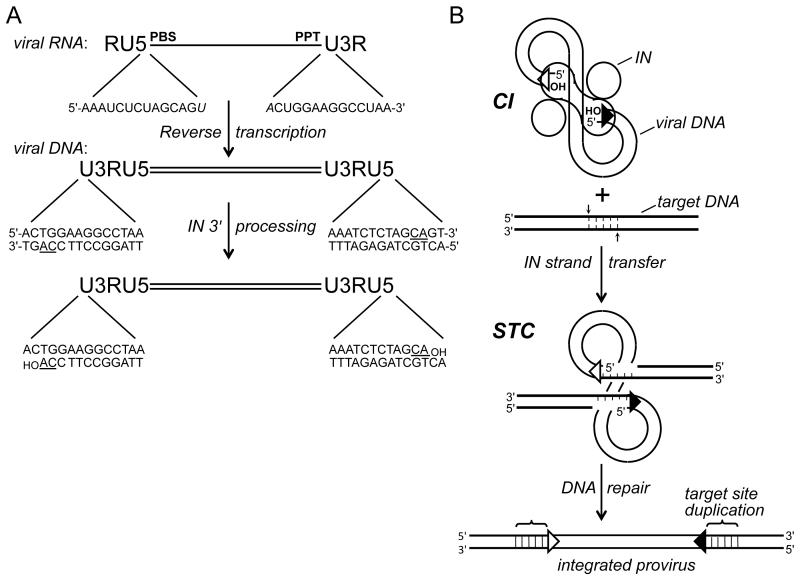
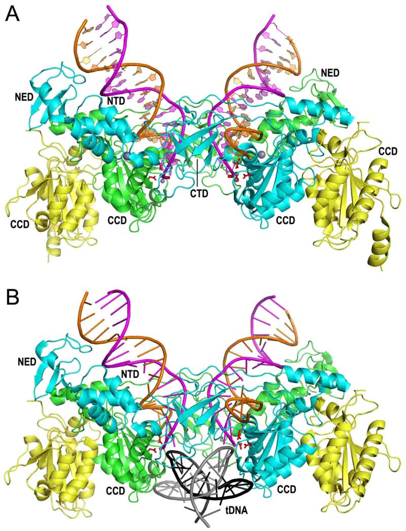
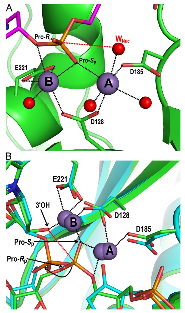
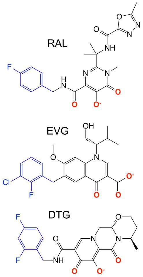
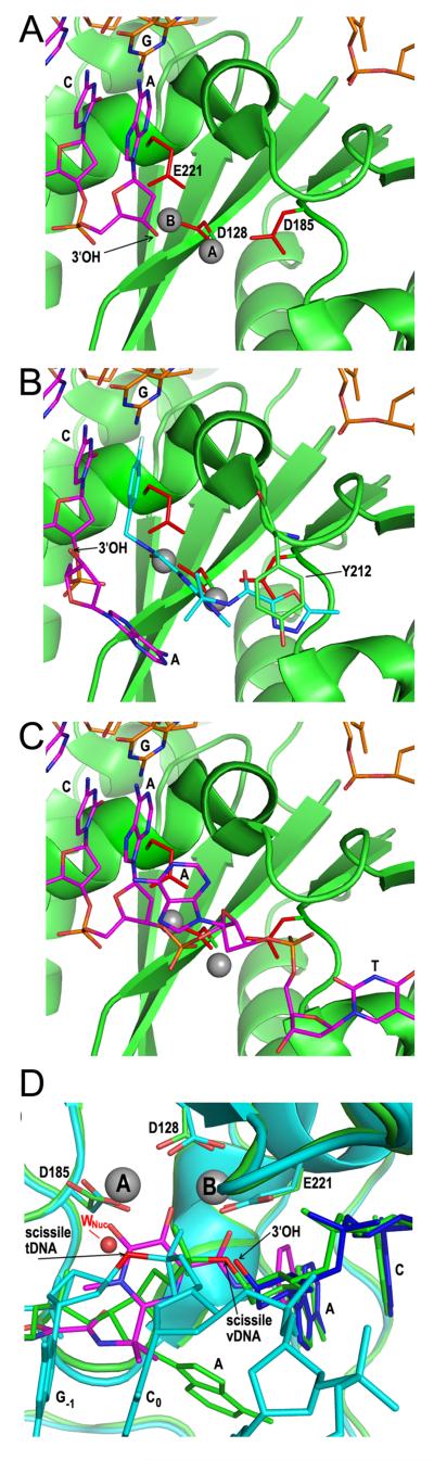
Due to the importance of human immunodeficiency virus type 1 (HIV-1) integrase as a drug target, the biochemistry and structural aspects of retroviral DNA integration have been the focus of intensive research during the past three decades. The retroviral integrase enzyme acts on the linear double-stranded viral DNA product of reverse transcription. Integrase cleaves specific phosphodiester bonds near the viral DNA ends during the 3' processing reaction. The enzyme then uses the resulting viral DNA 3'-OH groups during strand transfer to cut chromosomal target DNA, which simultaneously joins both viral DNA ends to target DNA 5'-phosphates. Both reactions proceed via direct transesterification of scissile phosphodiester bonds by attacking nucleophiles: a water molecule for 3' processing, and the viral DNA 3'-OH for strand transfer. X-ray crystal structures of prototype foamy virus integrase-DNA complexes revealed the architectures of the key nucleoprotein complexes that form sequentially during the integration process and explained the roles of active site metal ions in catalysis. X-ray crystallography furthermore elucidated the mechanism of action of HIV-1 integrase strand transfer inhibitors, which are currently used to treat AIDS patients, and provided valuable insights into the mechanisms of viral drug resistance.


Figures

Figure 1
Figure 1
Integration substrate and integrase activities. (A) Reverse transcription yields linear, double-stranded DNA with U3RU5 long terminal repeats. The 14 terminal bp of HIV-1 DNA are compared with their originating positions in viral RNA (italicized bases form part of the primer binding site (PBS) and polypurine tract (PPT) that are important for DNA synthesis). The positions of the invariant CA dinucleotides (underlined in the DNA) relative to the 3′ ends of the PBS and PPT determines whether 3′ processing is required to yield a CAOH 3′ terminus; 3′ processing by HIV-1 integrase liberates a pGpTOH dinucleotide from each viral DNA end. (B) Two monomers (oblong shape) of an HIV-1 integrase tetramer within the cleaved intasome (CI) use the viral DNA 3′-hydroxyl groups to cut the target DNA (thick bold lines) with a 5 bp stagger, which concomitantly joins the LTR ends to target DNA 5′ phosphates. Repair of the strand transfer complex (STC) yields a 5 bp duplication of target DNA flanking the integrated HIV-1 provirus. Open and filled triangles, U3 and U5 termini. Integrase was omitted from the drawing of the STC for simplicity. IN, integrase.
Figure 2
Figure 2
Retroviral integrase domain organization and HIV-1 integrase domain structures. (A) The N-terminal domain (NTD), catalytic core domain (CCD), and C-terminal domain (CTD) are common among all retroviral integrase proteins, whereas sequence analysis indicates that gammaretrovirus and epsilonretrovirus in addition to spumavirus proteins harbor an N-terminal extension domain (NED) (55, 94). HIV-1 and PFV integrases were aligned by NTD N-termini, with positions of domain boundaries and lengths of interdomain linker and C-terminal tail regions indicated. Residues conserved across all retroviral integrase proteins are shown in single letter code. Bars and arrows indicate alpha helix and beta strand secondary elements as determined by X-ray crystallography for PFV integrase (94) and by a combination of X-ray crystallography (66, 149) and molecular modeling (105) for HIV-1 integrase. (B) The X-ray crystal structure of the HIV-1 integrase CCD [protein database (pdb) code 1ITG] (43) highlights in red sticks the aspartate residues of the DDE catalytic triad (the glutamic acid was not visualized in this structure) and in blue sticks the Lys185 substitution that enhanced protein solubility and enabled protein crystallization (the Lys residue at the rear face of the dimer is barely visible in this projection). (C) The NMR structure of the integrase NTD (pdb code 1WJC) highlights the His (blue sticks) and Cys (yellow sticks) residues that coordinate a single zinc atom (grey sphere) (99). (D) The structure of the HIV-1 integrase CTD as determined by NMR (pdb code 1IHV) (142) highlights Arg231, which has been implicated in target DNA binding (108, 166).
Figure 3
Figure 3
Structures of 2-domain HIV-1 integrase constructs. (A) The X-ray crystal structure of the HIV-1 integrase CCD-CTD dimer (pdb code 1EX4) (66), highlighting the CCD and CTD side-chains that were shown in Fig. 2. (B) The crystal structure of the NTD-CCD asymmetric unit (pdb code 1K6Y) (149) highlights NTD residue Glu11 and CCD residue Lys186 of the green and cyan molecules, respectively, which play important roles in integrase concerted integration activity and HIV-1 infection (70). The other pair of interacting residues (Glu11 from the cyan NTD and Lys186 from the green CCD) is not visible in this projection. The side chains of the DDE catalytic triad (red sticks) and NTD-coordinated zinc atoms (grey spheres) are also shown.
Figure 4
Figure 4
Lentiviral integrase-LEDGF/p75 IBD structures. (A) X-ray crystal structure of the IBD (magenta cartoon)-HIV-1 integrase CCD/F185K (cyan/green dimer) complex (pdb code 2B4J) (69). Highlighted is the Asp366 side-chain from the upper IBD molecule (red stick) hydrogen bonding (dashed lines) to backbone amides of integrase residues within the linker between CCD α helices 4 and 5. The extent to which integrase CCD α4/5 connector regions contribute to forming analogous CCD-CCD dimer interface pockets at least in part accounts for the lentiviral specificity of the LEDGF/p75-integrase interaction (54, 69). The Asp and Glu side chains of the catalytic DDE triad are also painted red. (B) The crystal structure of the HIV-2 integrase NTD-CCD–IBD complex highlights the electrostatic interaction between the integrase NTD and the second helix-hairpin-helix unit of the IBD (pdb code 3F9K, chains A, B, and C) (61). Salt bridges between IBD residue Arg405 and integrase residue Glu10 are indicated by dashed lines; IBD residue Asp366 is behind the green CCD, hidden from view.
Figure 5
Figure 5
PFV intasome structures. (A) Structure containing 19 bp pre-cleaved U5 DNA end (94, 207) (pdb code 3OY9). The inner integrase monomers of the tetramer, which contact the viral DNA, are painted cyan and green; the outer integrase molecules are yellow. The transferred DNA strands with CAOH 3′ ends are painted magenta whereas the non-transferred strands are orange. The large grey spheres are NTD-coordinated zinc; small grey spheres are Mn atoms coordinated by the DDE active site residues (red sticks) and viral DNA end. (B) Structure of the TCC (98) (pdb code 3OS1). Although a 30 bp target DNA (tDNA) was utilized during crystallization, only 18 bp (grey plus strand sequence -7GCACGTG\CTAGCACGTGC10) was resolved in the electron density maps.
Figure 6
Figure 6
Structural basis of integrase 3′ processing and strand transfer activities. (A) Structure of the manganese-bound SSC (pdb code 4E7I). The DNA and integrase backbones are colored magenta and green, respectively; red and orange sticks are oxygen and phosphorus atoms, respectively. Gray and red spheres are manganese ions and water molecules, respectively, with the nucleophilic water labeled WNuc. Black dashed lines indicate metal ion interactions; the red dashed line connects the nucleophile and scissile phosphodiester bond. (B) Overlay of metal ion-bound TCC (pdb code 4E7K; DNA and protein in green) and STC (pdb code 4E7L; elements painted in cyan) structures. Both sets of metal ions are shown; the 3.8 Å spacing between ions in the TCC contracts to 3.2 Å in the STC (85). The curved black line indicates the displacement of the viral-target DNA phosphodiester bond after strand transfer relative to the scissile bond in target DNA. Other labeling is the same as in panel A.
Figure 7
Figure 7
Chemical structures of INSTIs raltegravir (RAL), elvitegravir (EVG), and dolutegravir (DTG).
Figure 8
Figure 8
Structural basis of INSTI mechanism of action. (A) The active site from pdb code 3OY9 highlights PFV integrase DDE residues, Mn2+ ions A and B, and the 3′-OH of the terminal deoxyadenylate. (B) Structure of raltegravir (cyan)-bound integrase active site (pdb code 3OYA) highlighting positions of supplanted deoxyadenylate 3′-OH, magnesium ions (grey), and integrase residue Tyr212. (C) The integrase active site in the context of the PFV SSC highlights the position of the unprocessed AT dinucleotide. Additional panel A-C coloring: green, integrase; magenta, transferred DNA strand; orange, non-transferred strand. (D) Overlaid structures of the PFV SSC (pdb code 4E7I; integrase and viral DNA in green), TCC (pdb code 4E7K; integrase and target DNA in cyan and viral DNA in blue), and raltegravir-bound CI (pdb code 3OYA; raltegravir in magenta) highlights the common positioning of raltegravir oxygen atoms with strand transfer and 3′ processing attacking and leaving groups. Subscript numbers denote target DNA bases. tDNA, target DNA; vDNA, viral DNA.

References

    1. Métifiot M, Marchand C, Pommier Y. HIV integrase inhibitors: 20-year landmark and challenges. Adv. Pharmacol. 2013;67:75–105. - PMC - PubMed
    1. Ruelas DS, Greene WC. An integrated overview of HIV-1 latency. Cell. 2013;155:519–529. - PMC - PubMed
    1. Donehower LA, Varmus HE. A mutant murine leukemia virus with a single missense codon in pol is defective in a function affecting integration. Proc. Natl. Acad. Sci. USA. 1984;81:6461–6465. - PMC - PubMed
    1. Panganiban AT, Temin HM. The retrovirus pol gene encodes a product required for DNA integration: Identification of a retrovirus int locus. Proc. Natl. Acad. Sci. USA. 1984;81:7885–7889. - PMC - PubMed
    1. Schwartzberg P, Colicelli J, Goff SP. Construction and analysis of deletion mutations in the pol gene of moloney murine leukemia virus: A new viral function required for productive infection. Cell. 1984;37:1043–1052. - PubMed

LinkOut - more resources