. 2015:2015:729831.
doi: 10.1155/2015/729831. Epub 2015 Feb 1.

Thaliporphine derivative improves acute lung injury after traumatic brain injury

Affiliations

Thaliporphine derivative improves acute lung injury after traumatic brain injury

Gunng-Shinng Chen et al. Biomed Res Int. 2015.

Abstract

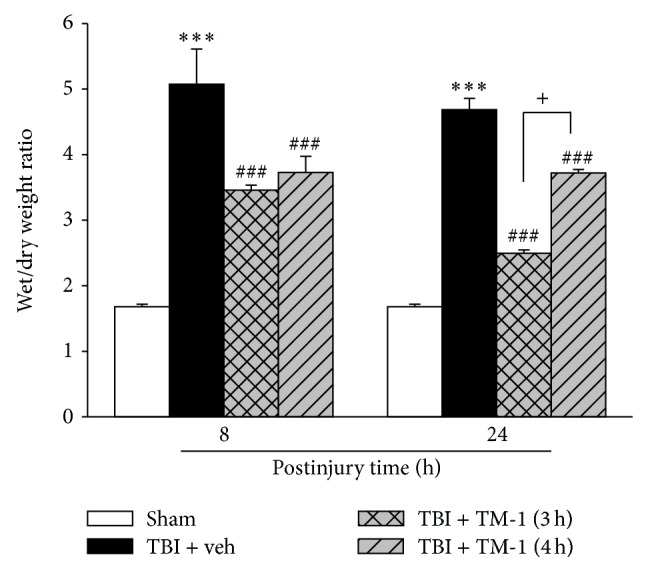
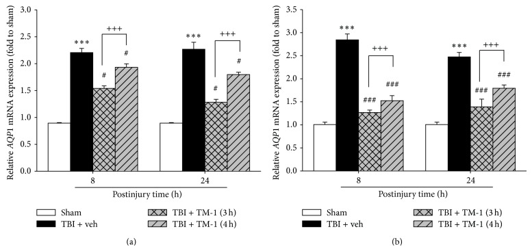
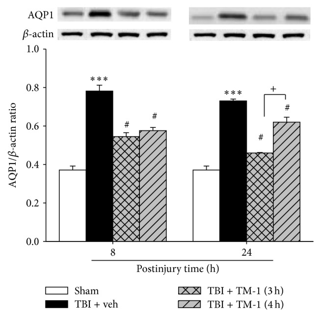
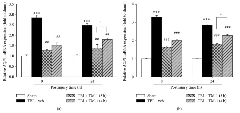
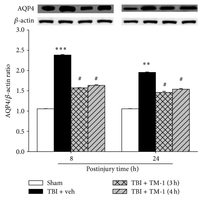
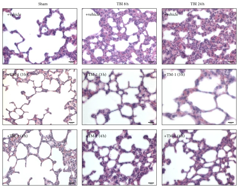
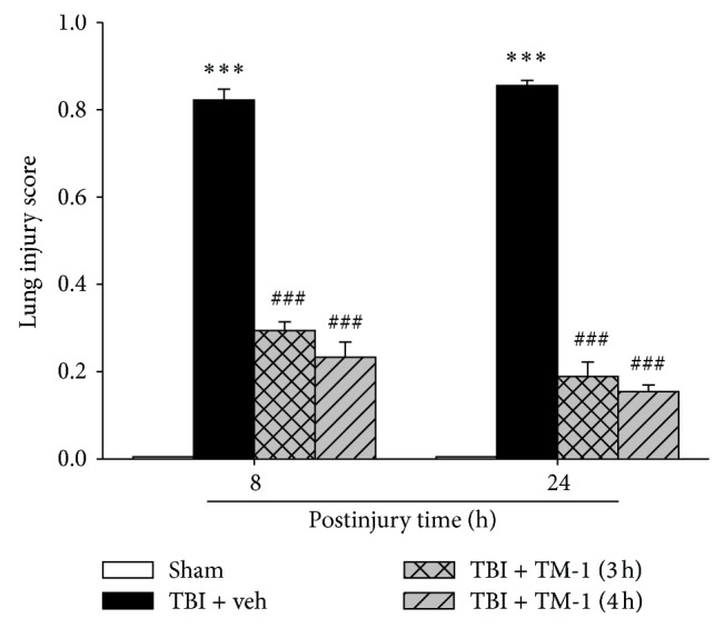
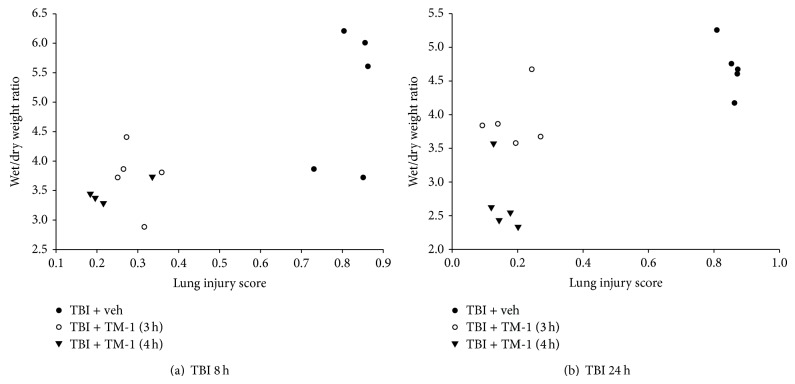
Acute lung injury (ALI) occurs frequently in patients with severe traumatic brain injury (TBI) and is associated with a poor clinical outcome. Aquaporins (AQPs), particularly AQP1 and AQP4, maintain water balances between the epithelial and microvascular domains of the lung. Since pulmonary edema (PE) usually occurs in the TBI-induced ALI patients, we investigated the effects of a thaliporphine derivative, TM-1, on the expression of AQPs and histological outcomes in the lung following TBI in rats. TM-1 administered (10 mg/kg, intraperitoneal injection) at 3 or 4 h after TBI significantly reduced the elevated mRNA expression and protein levels of AQP1 and AQP4 and diminished the wet/dry weight ratio, which reflects PE, in the lung at 8 and 24 h after TBI. Postinjury TM-1 administration also improved histopathological changes at 8 and 24 h after TBI. PE was accompanied with tissue pathological changes because a positive correlation between the lung injury score and the wet/dry weight ratio in the same animal was observed. Postinjury administration of TM-1 improved ALI and reduced PE at 8 and 24 h following TBI. The pulmonary-protective effect of TM-1 may be attributed to, at least in part, downregulation of AQP1 and AQP4 expression after TBI.


Figures

Figure 1
Figure 1
TM-1 administered 3 or 4 h after injury reduced the TBI-induced lung wet/dry weight ratio measured 8 and 24 h after TBI. Decrease in the wet/dry weight ratio at 3 or 4 h after injury with TM-1 administration (10 mg/kg) when the rats were sacrificed at 8 or 24 h after injury, respectively. Data are expressed as mean ± SD (n = 5/group). *** P < 0.001 TBI + veh group compared with the sham-operated group; ### P < 0.001 TM-1 treated groups compared with the TBI + veh groups; + P < 0.05, comparison between wet/dry weight ratios of rats sacrificed at 24 h following TM-1 administration (10 mg/kg) at 3 and 4 h after injury.
Figure 2
Figure 2
TM-1 administered 3 or 4 h after injury reduced the TBI-induced AQP1 mRNA expression measured 8 and 24 h after TBI. Relative AQP1 mRNA expression was measured by reverse transcription Q-PCR using β-actin (a) and Gapdh (b) as reference genes. AQP1 mRNA expression significantly decreased following TM-1 administration (10 mg/kg) at 3 and 4 h after injury in the rats sacrificed 8 or 24 h after injury, respectively. Compared with the TBI + veh groups, the TM-1 treated groups exhibited significantly decreased mRNA expression. Data are expressed as mean ± SD (n = 5/group). *** P < 0.001 TBI + veh group compared with the sham-operated group; # P < 0.05 and ### P < 0.0001 TM-1 treated group compared with the TBI + veh groups; +++ P < 0.001, comparison between mRNA expression following TM-1 administration (10 mg/kg) 3 and 4 h after injury in the rats sacrificed 8 or 24 h after TBI.
Figure 3
Figure 3
TM-1 administered 3 or 4 h after injury reduced TBI-induced lung AQP1 protein expression levels measured 8 and 24 h after TBI. The AQP1 protein level showed a marked decrease following TM-1 administration 3 or 4 h after injury (10 mg/kg), respectively. Rats were sacrificed 8 or 24 h after TBI. Data are expressed as mean ± SD (n = 5/group). *** P < 0.001 compared with the sham-operated group; # P < 0.05 compared with the untreated groups; +++ P < 0.001, comparison between mRNA expression following TM-1 administration (10 mg/kg) 3 and 4 h after injury in the rats sacrificed 8 and 24 h after TBI; + P < 0.05, comparison between protein levels following TM-1 administration (10 mg/kg) 3 and 4 h after injury in the rats sacrificed 24 h after TBI.
Figure 4
Figure 4
TM-1 administered 3 or 4 h after injury reduced the TBI-induced AQP4 mRNA expression measured 8 and 24 h after TBI. (a) β-actin and (b) Gapdh as reference genes. AQP4 mRNA expression significantly decreased following TM-1 administration (10 mg/kg) 3 and 4 h after injury in the rats sacrificed 8 or 24 h after injury, respectively. Compared with the TBI + veh groups, the TM-1 treated groups exhibited significantly decreased mRNA expression. Data are expressed as mean ± SD (n = 5/group). *** P < 0.001 TBI + veh group compared with the sham-operated group; ## P < 0.01 and ### P < 0.001 compared with the TBI + veh groups; + P < 0.05, comparison between mRNA expression following TM-1 administration (10 mg/kg) 3 and 4 h after injury in the rats sacrificed 8 or 24 h after TBI.
Figure 5
Figure 5
TM-1 administered 3 or 4 h after injury reduced the TBI-induced AQP4 protein level measured 8 and 24 h after TBI. The AQP4 protein level showed a marked decrease following TM-1 administration (10 mg/kg) 3 and 4 h after injury, respectively. Rats were sacrificed 8 or 24 h after TBI. Data are expressed as mean ± SD (n = 5/group). ** P < 0.01 and *** P < 0.001 TBI + veh group compared with the sham-operated group; # P < 0.05 TM-1 treated groups compared with the TBI + veh groups; no significance in AQP4 protein expression following TM-1 administration (10 mg/kg) at 3 and 4 h after injury in the rats sacrificed at 8 or 24 h after TBI.
Figure 6
Figure 6
TM-1 administered 3 or 4 h after injury reduced alveolar swelling with infiltration of inflammatory cells 8 and 24 h after TBI by H&E staining. The vehicle-treated sham-operated group showed a normal alveolar morphology. Alveolar swelling and infiltration of inflammatory cells increased 8 and 24 h in vehicle-treated TBI rats. Alveolar swelling and infiltration of inflammatory cells decreased following TM-1 administration 3 and 4 h after injury. Rats were sacrificed 8 or 24 h after TBI. Bar = 25 μm.
Figure 7
Figure 7
TM-1 administered 3 or 4 h after injury reduced the TBI-induced lung injury score 8 and 24 h after TBI. Significant difference was observed between the sham-operated and TBI groups. The lung injury score decreased with TM-1 administration (10 mg/kg) 3 and 4 h after injury. Rats were sacrificed 24 h after TBI. The lung injury score decreased following TM-1 administration (10 mg/kg) 3 and 4 h after injury. Rats were sacrificed 24 h after TBI. Data are expressed as mean ± SD (n = 5/group). *** P < 0.001 compared with the sham-operated group; ### P < 0.001 compared with the untreated groups.
Figure 8
Figure 8
Rats administered TM-1 3 or 4 h after injury showed a positive correlation between the wet/dry weight ratio and the lung injury score. (a) The wet/dry weight ratio and the lung injury score in rats sacrificed at 8 h after TBI exhibited a positive correlation (df = 13, r = 0.72224) and a significant difference (P < 0.01) in the three groups. (b) The wet/dry weight ratio and the lung injury score in rats sacrificed at 24 h after TBI exhibited a positive correlation (df = 13, r = 0.72633) and a significant difference (P < 0.01) in the three groups.

References

    1. Baumann A., Audibert G., McDonnell J., Mertes P. M. Neurogenic pulmonary edema. Acta Anaesthesiologica Scandinavica. 2007;51(4):447–455. doi: 10.1111/j.1399-6576.2007.01276.x. - DOI - PubMed
    1. Gajic O., Manno E. M. Neurogenic pulmonary edema: another multiple-hit model of acute lung injury. Critical Care Medicine. 2007;35(8):1979–1980. doi: 10.1097/01.CCM.0000277254.12230.7D. - DOI - PubMed
    1. Zygun D. A., Kortbeek J. B., Fick G. H., Laupland K. B., Doig C. J. Non-neurologic organ dysfunction in severe traumatic brain injury. Critical Care Medicine. 2005;33(3):654–660. doi: 10.1097/01.CCM.0000155911.01844.54. - DOI - PubMed
    1. Martin G. S., Brigham K. L. Fluid flux and clearance in acute lung injury. Comprehensive Physiology. 2012;2(4):2471–2480. doi: 10.1002/cphy.c100050. - DOI - PubMed
    1. Kandatsu N., Nan Y.-S., Feng Q.-G., et al. Opposing effects of isoflurane and sevoflurane on neurogenic pulmonary edema development in an animal model. Anesthesiology. 2005;102(6):1182–1189. doi: 10.1097/00000542-200506000-00018. - DOI - PubMed

Publication types