

Anterior Mediastinal Masses in the Framingham Heart Study: Prevalence and CT Image Characteristics
- PMID: 25705709
- PMCID: PMC4332399
- DOI: 10.1016/j.ejro.2014.12.003
Anterior Mediastinal Masses in the Framingham Heart Study: Prevalence and CT Image Characteristics
Abstract
Purpose: To investigate the prevalence and CT image characteristics of anterior mediastinal masses in a population-based cohort and their association with the demographics of the participants.
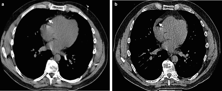
Materials and methods: Chest CT scans of 2571 Framingham Heart Study participants (mean age 58.9 years, 51% female) were evaluated by two board-certified radiologists with expertise in thoracic imaging for the presence of anterior mediastinal masses, their shape, contour, location, invasion of adjacent structures, fat content, and calcification. For participants with anterior mediastinal masses, a previous cardiac CT scan was reviewed for interval size change of the masses, when available. The demographics of the participants were studied for any association with the presence of anterior mediastinal masses.
Results: Of 2571, 23 participants (0.9%, 95% CI: 0.6 to 1.3) had anterior mediastinal masses on CT. The most common CT characteristics were oval shape, lobular contour, and midline location, showing soft tissue density (median 32.1 HU). Fat content was detected in a few cases (9%, 2/23). Six out of eight masses with available prior cardiac CT scans demonstrated an interval growth over a median period of 6.5 years. No risk factors for anterior mediastinal masses were detected among participants' demographics, including age, sex, BMI, and cigarette smoking.
Conclusions: The prevalence of anterior mediastinal masses is 0.9% in the Framingham Heart Study. Those masses may increase in size when observed over 5-7 years. Investigation of clinical significance in incidentally found anterior mediastinal masses with a longer period of follow-up would be necessary.
Keywords: Anterior mediastinal masses; CT; Prevalence; the Framingham Heart Study.
Figures


References
-
- Tomiyama N., Honda O., Tsubamoto M., Inoue A., Sumikawa H., Kuriyama K. Anterior mediastinal tumors: diagnostic accuracy of CT and MRI. Eur J Radiol. 2009;69(2):280–288. - PubMed
-
- Henschke C.I., Lee I.J., Wu N., Farooqi A., Khan A., Yankelevitz D. CT screening for lung cancer: prevalence and incidence of mediastinal masses. Radiology. 2006;239(2):586–590. - PubMed
-
- Baron R., Lee J., Sagel S., Levitt R. Computed tomography of the abnormal thymus. Radiology. 1982;142(1):127–134. - PubMed
-
- Inaoka T., Takahashi K., Mineta M., Yamada T., Shuke N., Okizaki A. Thymic hyperplasia and thymus gland tumors: differentiation with chemical shift MR imaging. Radiology. 2007;243(3):869–876. - PubMed
Grants and funding
LinkOut - more resources
Full Text Sources
Other Literature Sources
