

X-linked macrocytic dyserythropoietic anemia in females with an ALAS2 mutation
- PMID: 25705881
- PMCID: PMC4396476
- DOI: 10.1172/JCI78619
X-linked macrocytic dyserythropoietic anemia in females with an ALAS2 mutation
Erratum in
-
X-linked macrocytic dyserythropoietic anemia in females with an ALAS2 mutation.J Clin Invest. 2020 Jan 2;130(1):552. doi: 10.1172/JCI132538. J Clin Invest. 2020. PMID: 31895053 Free PMC article. No abstract available.
Abstract
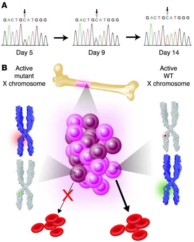
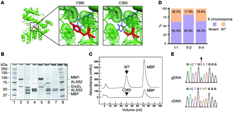
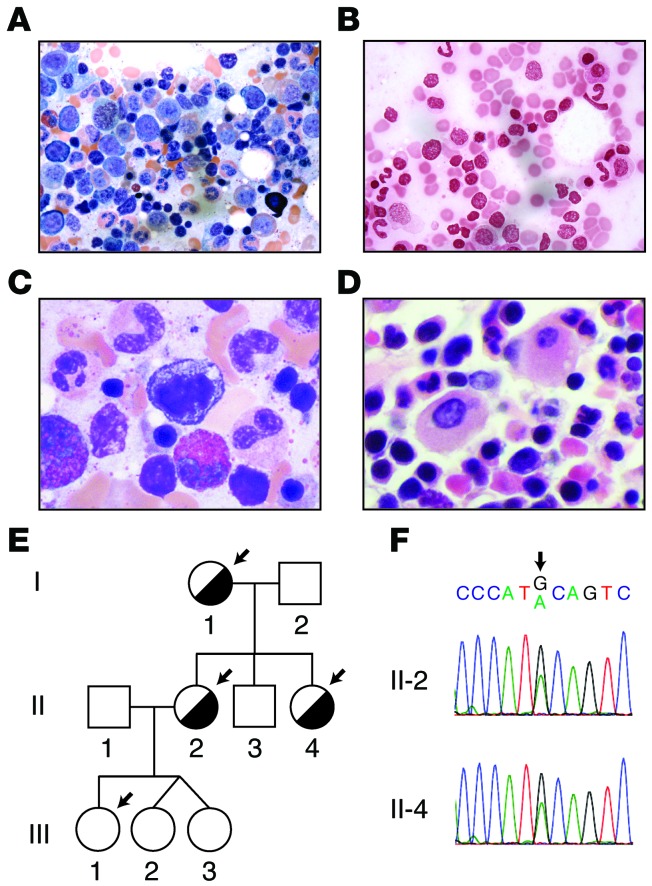
Macrocytic anemia with abnormal erythropoiesis is a common feature of megaloblastic anemias, congenital dyserythropoietic anemias, and myelodysplastic syndromes. Here, we characterized a family with multiple female individuals who have macrocytic anemia. The proband was noted to have dyserythropoiesis and iron overload. After an extensive diagnostic evaluation that did not provide insight into the cause of the disease, whole-exome sequencing of multiple family members revealed the presence of a mutation in the X chromosomal gene ALAS2, which encodes 5'-aminolevulinate synthase 2, in the affected females. We determined that this mutation (Y365C) impairs binding of the essential cofactor pyridoxal 5'-phosphate to ALAS2, resulting in destabilization of the enzyme and consequent loss of function. X inactivation was not highly skewed in wbc from the affected individuals. In contrast, and consistent with the severity of the ALAS2 mutation, there was a complete skewing toward expression of the WT allele in mRNA from reticulocytes that could be recapitulated in primary erythroid cultures. Together, the results of the X inactivation and mRNA studies illustrate how this X-linked dominant mutation in ALAS2 can perturb normal erythropoiesis through cell-nonautonomous effects. Moreover, our findings highlight the value of whole-exome sequencing in diagnostically challenging cases for the identification of disease etiology and extension of the known phenotypic spectrum of disease.
Figures
Similar articles
-
A Novel ALAS2 Mutation Resulting in Variable Phenotypes and Pyridoxine Response in a Family with X-linked Sideroblastic Anemia.Ann Clin Lab Sci. 2017 May;47(3):319-322. Ann Clin Lab Sci. 2017. PMID: 28667034
-
Identification of a novel heterozygous ALAS2 mutation in a young Chinese female with X-linked sideroblastic anemia.Ann Hematol. 2020 Feb;99(2):371-373. doi: 10.1007/s00277-019-03894-6. Epub 2019 Dec 17. Ann Hematol. 2020. PMID: 31848684 No abstract available.
-
Onset of X-linked sideroblastic anemia in the fourth decade.Haematologica. 2004 Oct;89(10):1261-3. Haematologica. 2004. PMID: 15477213
-
[Congenital sideroblastic anemia-a new family with identification of K156E mutation of ALAS2 gene and literature review].Zhonghua Xue Ye Xue Za Zhi. 2014 Feb;35(2):142-6. doi: 10.3760/cma.j.issn.0253-2727.2014.02.018. Zhonghua Xue Ye Xue Za Zhi. 2014. PMID: 24606657 Review. Chinese.
-
Molecular defects of erythroid 5-aminolevulinate synthase in X-linked sideroblastic anemia.J Bioenerg Biomembr. 1995 Apr;27(2):161-8. doi: 10.1007/BF02110031. J Bioenerg Biomembr. 1995. PMID: 7592563 Review.
Cited by
-
Understanding Sideroblastic Anemia: An Overview of Genetics, Epidemiology, Pathophysiology and Current Therapeutic Options.J Blood Med. 2020 Sep 25;11:305-318. doi: 10.2147/JBM.S232644. eCollection 2020. J Blood Med. 2020. PMID: 33061728 Free PMC article. Review.
-
Azacitidine is a potential therapeutic drug for pyridoxine-refractory female X-linked sideroblastic anemia.Blood Adv. 2022 Feb 22;6(4):1100-1114. doi: 10.1182/bloodadvances.2021005664. Blood Adv. 2022. PMID: 34781359 Free PMC article.
-
[New mutation of congenital sideroblastic anemia: a case report and literature review].Zhonghua Xue Ye Xue Za Zhi. 2021 Jul 14;42(7):603-605. doi: 10.3760/cma.j.issn.0253-2727.2021.07.013. Zhonghua Xue Ye Xue Za Zhi. 2021. PMID: 34455750 Free PMC article. Review. Chinese. No abstract available.
-
X-linked sideroblastic anaemia in a female fetus: a case report and a literature review.BMC Med Genomics. 2021 Dec 20;14(1):296. doi: 10.1186/s12920-021-01146-z. BMC Med Genomics. 2021. PMID: 34930268 Free PMC article. Review.
-
Functional Selectivity in Cytokine Signaling Revealed Through a Pathogenic EPO Mutation.Cell. 2017 Mar 9;168(6):1053-1064.e15. doi: 10.1016/j.cell.2017.02.026. Cell. 2017. PMID: 28283061 Free PMC article.
References
-
- Davenport J. Macrocytic anemia. Am Fam Physician. 1996;53(1):155–162. - PubMed
