Case Reports
. 2015 Apr;125(4):1670-8.
doi: 10.1172/JCI79765. Epub 2015 Feb 23.

Identification of a human synaptotagmin-1 mutation that perturbs synaptic vesicle cycling

Case Reports

Identification of a human synaptotagmin-1 mutation that perturbs synaptic vesicle cycling

Kate Baker et al. J Clin Invest. 2015 Apr.

Abstract

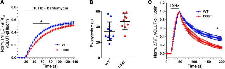
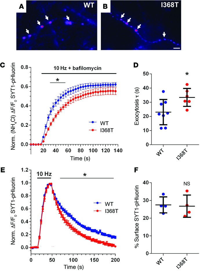
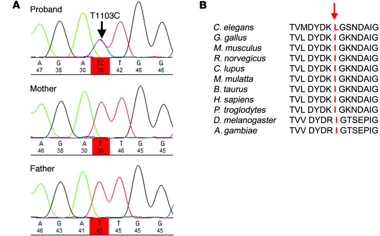
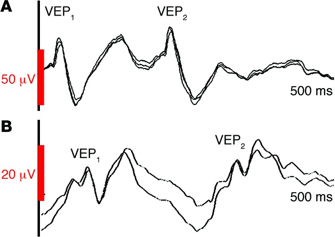
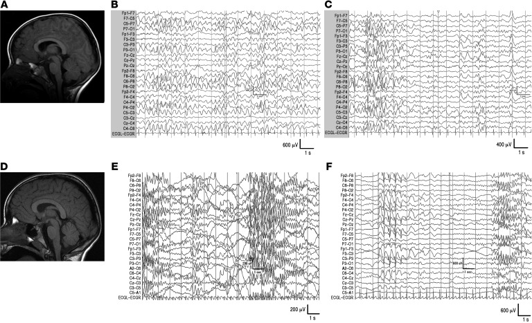
Synaptotagmin-1 (SYT1) is a calcium-binding synaptic vesicle protein that is required for both exocytosis and endocytosis. Here, we describe a human condition associated with a rare variant in SYT1. The individual harboring this variant presented with an early onset dyskinetic movement disorder, severe motor delay, and profound cognitive impairment. Structural MRI was normal, but EEG showed extensive neurophysiological disturbances that included the unusual features of low-frequency oscillatory bursts and enhanced paired-pulse depression of visual evoked potentials. Trio analysis of whole-exome sequence identified a de novo SYT1 missense variant (I368T). Expression of rat SYT1 containing the equivalent human variant in WT mouse primary hippocampal cultures revealed that the mutant form of SYT1 correctly localizes to nerve terminals and is expressed at levels that are approximately equal to levels of endogenous WT protein. The presence of the mutant SYT1 slowed synaptic vesicle fusion kinetics, a finding that agrees with the previously demonstrated role for I368 in calcium-dependent membrane penetration. Expression of the I368T variant also altered the kinetics of synaptic vesicle endocytosis. Together, the clinical features, electrophysiological phenotype, and in vitro neuronal phenotype associated with this dominant negative SYT1 mutation highlight presynaptic mechanisms that mediate human motor control and cognitive development.


Figures

Figure 5
Figure 5. Effect of I368T mutation on activity-dependent SV cycling.
Neurons were cotransfected with vGLUT-pHluorin and either mCer-SYT1WT (WT) or mCer-SYT1I368T (I368T). (A and B) Neurons were stimulated with a train of 1,200 action potentials at 10 Hz in the presence of bafilomycin as described for Figure 4C. (A) Average time course of ΔF/F0 for vGLUT-pHluorin ± SEM, normalized to NH4Cl (n = 11 WT, n = 9 I368T. *P < 0.05, over time indicated (2-way ANOVA). (B) Average exocytosis time constant (τ) determined by fitting single-phase association curve to data presented in A. Data are presented as mean ± SEM (n = 11 WT, n = 9 I368T). *P < 0.02 (t test). (C) Neurons were stimulated with a train of 300 action potentials at 10 Hz. Graph displays the average time course of ΔF/F0 of vGLUT-pHluorin ± SEM, normalized to peak of stimulation (n = 8 WT, n = 9 I368T). *P < 0.05, over time indicated (2-way ANOVA).
Figure 4
Figure 4. Effect of I368T mutation on SYT1 localization and activity-dependent trafficking.
(A and B) Hippocampal neurons were transfected with either SYT1WT-pHluorin or SYT1I368T-pHluorin. Representative images show a similar punctate distribution of SYT1WT-pHluorin (WT, A) and SYT1I368T-pHluorin (I368T, B) along axonal segments after exposure to alkaline imaging buffer. Image is displayed in false color, with warmer colors indicating more fluorescence intensity. Arrows represent the localization of both pHluorin reporters at nerve terminals. Scale bar: 1 μm. (C and D) Transfected neurons were stimulated with a train of 1,200 action potentials at 10 Hz in the presence of 1 μM bafilomycin to prevent vesicle reacidification and subsequently exposed to NH4Cl to reveal total pHluorin fluorescence (n = 8 WT, n = 6 I368T). (C) Time course of mean ΔF/F0 of SYT1-pHluorin ± SEM, normalized to NH4Cl. *P < 0.05, over time indicated (2-way ANOVA). (D) Exocytosis time constant (τ) determined by fitting a single-phase association curve to the data presented in C. Data are presented as mean ± SEM (n = 8 WT and n = 6 I368T). *P < 0.03 (t test). (E) Neurons were stimulated with a train of 300 action potentials at 10 Hz. Graph displays the time course of mean ΔF/F0 of SYT1-pHluorin ± SEM, normalized to the peak of stimulation (n = 7 WT, n = 6 I368T). *P < 0.05, over time indicated (2-way ANOVA). (F) Surface SYT1-pHluorin fluorescence, determined by normalizing SYT1-pHluorin fluorescence to 100% in alkaline buffer and 0% in acidic buffer. Data are presented as mean ± SEM. n = 4. P = 0.86 (t test).
Figure 3
Figure 3. Identification, confirmation, and evolutionary analysis of de novo SYT1 variant.
(A) Sanger sequence analysis confirming the de novo SYT1 variant. Sequence chromatograms demonstrate the presence of the variant in the proband and the reference allele in both parents. (B) Alignment of cross-species SYT1 protein sequences, showing the position of the mutation I368T at the absolutely conserved isoleucine residue within a highly conserved region of amino acid sequence.
Figure 2
Figure 2. VEPs in proband with de novo heterozygous SYT1 I368T mutation and age-matched control.
Paired-pulse flash stimulation at IPI of 200 ms, sweep length 500 ms. (A) Healthy 8-year-old child: amplitude of VEP2 is at least 80% of the amplitude of VEP1. (B) Proband: amplitude of VEP2 waveform is around 50% of the amplitude of VEP1. Note difference in VEP1 amplitude and latency between the proband and control. See also Supplemental Figure 2 (auditory evoked potentials).
Figure 1
Figure 1. MRI and EEG in proband with de novo heterozygous SYT1 I368T mutation.
(A) MRI at 2 years; (B) EEG at 2 years (awake); (C) EEG at 2 years (asleep); (D) MRI at 8 years; (E) EEG at 8 years (awake); (F) EEG at 8 years (asleep). T1-weighted MR images (midline sagittal slices) are presented in radiological orientation. See also Supplemental Figure 1 (spectral analysis).

References

    1. Zoghbi HY, Bear MF. Synaptic dysfunction in neurodevelopmental disorders associated with autism and intellectual disabilities. Cold Spring Harb Perspect Biol. 2012;4(3):a009886. - PMC - PubMed
    1. Waites CL, Garner CC. Presynaptic function in health and disease. Trends Neurosci. 2011;34(6):326–337. doi: 10.1016/j.tins.2011.03.004. - DOI - PubMed
    1. Südhof TC, Rizo J. Synaptic vesicle exocytosis. Cold Spring Harb Perspect Biol. 2011;3(12):a005637. - PMC - PubMed
    1. Geppert M, et al. Synaptotagmin I: a major Ca2+ sensor for transmitter release at a central synapse. Cell. 1994;79(4):717–727. doi: 10.1016/0092-8674(94)90556-8. - DOI - PubMed
    1. Bacaj T, et al. Synaptotagmin-1 and synaptotagmin-7 trigger synchronous and asynchronous phases of neurotransmitter release. Neuron. 2013;80(4):947–959. doi: 10.1016/j.neuron.2013.10.026. - DOI - PMC - PubMed

MeSH terms