. 2015 Feb 23;10(2):e0116086.
doi: 10.1371/journal.pone.0116086. eCollection 2015.

A distinct mechanism of vascular lumen formation in Xenopus requires EGFL7

Affiliations

A distinct mechanism of vascular lumen formation in Xenopus requires EGFL7

Marta S Charpentier et al. PLoS One. .

Abstract

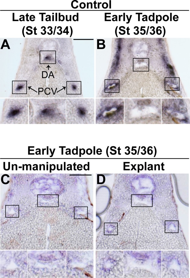
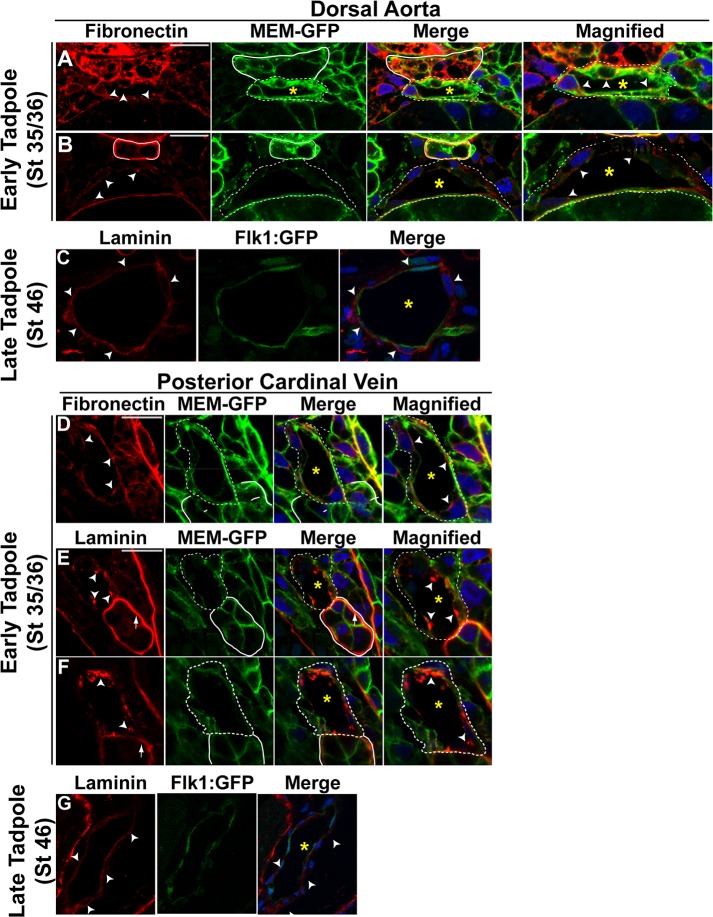
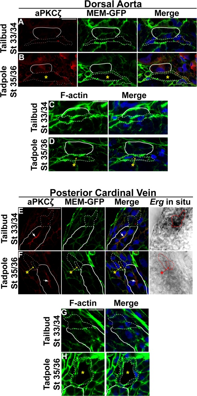
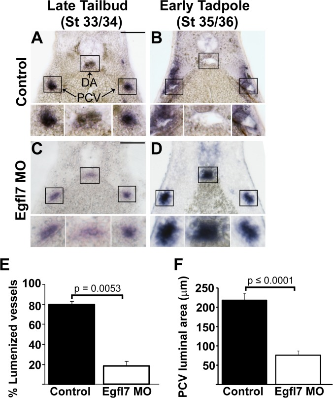
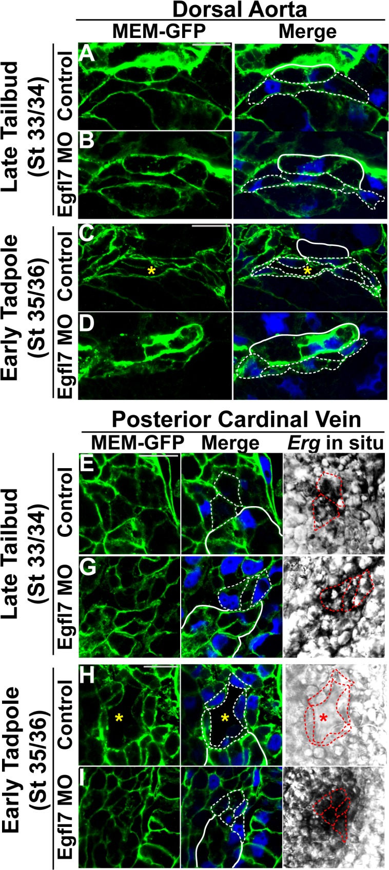
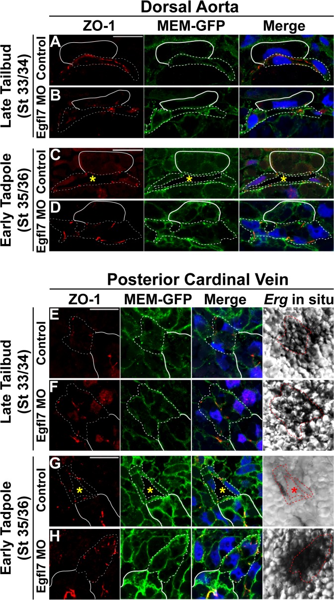
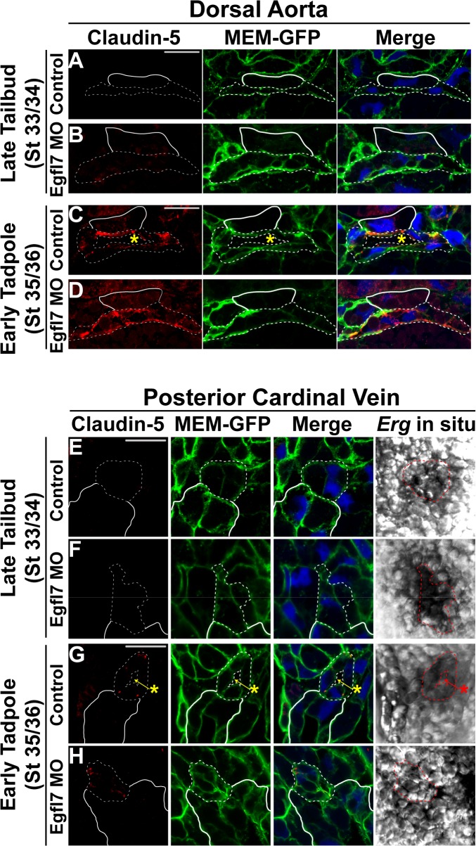
During vertebrate blood vessel development, lumen formation is the critical process by which cords of endothelial cells transition into functional tubular vessels. Here, we use Xenopus embryos to explore the cellular and molecular mechanisms underlying lumen formation of the dorsal aorta and the posterior cardinal veins, the primary major vessels that arise via vasculogenesis within the first 48 hours of life. We demonstrate that endothelial cells are initially found in close association with one another through the formation of tight junctions expressing ZO-1. The emergence of vascular lumens is characterized by elongation of endothelial cell shape, reorganization of junctions away from the cord center to the periphery of the vessel, and onset of Claudin-5 expression within tight junctions. Furthermore, unlike most vertebrate vessels that exhibit specialized apical and basal domains, we show that early Xenopus vessels are not polarized. Moreover, we demonstrate that in embryos depleted of the extracellular matrix factor Epidermal Growth Factor-Like Domain 7 (EGFL7), an evolutionarily conserved factor associated with vertebrate vessel development, vascular lumens fail to form. While Claudin-5 localizes to endothelial tight junctions of EGFL7-depleted embryos in a timely manner, endothelial cells of the aorta and veins fail to undergo appropriate cell shape changes or clear junctions from the cell-cell contact. Taken together, we demonstrate for the first time the mechanisms by which lumens are generated within the major vessels in Xenopus and implicate EGFL7 in modulating cell shape and cell-cell junctions to drive proper lumen morphogenesis.


Conflict of interest statement

Competing Interests: The authors have declared that no competing interests exist.

Figures

Fig 1
Fig 1. Vascular lumen formation proceeds independently of circulation in Xenopus.
(A-B). In situ hybridization with endothelial-specific Erg followed by sectioning was performed to determine the time course of vascular lumen formation of the dorsal aorta (DA) and posterior cardinal veins (PCV) in Xenopus. (A). Endothelial cells are coalesced together at the correct positions of the DA and PCVs in late tailbud stage (stage 33/34) control embryos. (B). Lumenized DA and PCVs become apparent by early tadpole stage (stage 35/36) in control embryos. 3–4 embryos from each condition/stage were assessed from at least three independent injection batches at the same position along the anterior-posterior axis of the embryo. (C-D). In situ hybridization with Erg followed by sectioning. (C). Transverse section of an early tadpole stage (stage 35/36) wildtype embryo depicting lumenized DA and PCVs. (D). Following the removal of the anterior and heart of early tailbud embryos, explants were cultured until early tadpole stage (stage 35/36) and processed as indicated above. PCV and DA lumens readily form in the absence of a functioning heart or circulatory system. 3–4 embryos from each condition were assessed from two independent experiments. Top image of each panel was taken at 20x magnification. Black boxes correspond to enlarged images of each vessel displayed below. Scale bars represent 20 μm.
Fig 2
Fig 2. Basement membrane proteins are deposited on both the basal and apical surfaces of early vessels in Xenopus.
(A-G) Representative confocal images of transverse sections of the dorsal aorta (DA) and posterior cardinal veins (PCV) stained with laminin or fibronectin (red). In A,B,D,E,F staining with GFP to mark cell membranes (MEM-GFP) and DAPI to mark nuclei (blue) is also shown. In C and G, Flk-1:GFP transgenic animals were used to identify endothelial cells (green). All images taken at 63x magnification with scale bars indicating 20 μm. Dotted white lines in all panels delineate the endothelial cells comprising each vessel. As anatomical references, the hypochord in (A-B) and the kidney in (D-F) are indicated by solid white lines. Dotted lines in the magnified images represent vessels. Asterisks denote the vessel lumen. (A). The DA of early tadpole stage (stage 35/36) control embryos display deposition of fibronectin on both apical and basal surfaces (arrowheads point to apical staining). (B). DA of early tadpole stage (stage 35/36) control embryo exhibits the laminin staining on the basal and the apical surfaces of the vessel (arrowheads point to apical staining). (C). By late tadpole stage (stage 46), proper polarity is established in the dorsal aorta with laminin localizing to the basal surface of endothelial cells (arrowheads point to basal staining). (D) PCV of early tadpole stage (stage 35/36) control embryo exhibits apically and basally deposited fibronectin (arrowheads point to apical staining). (E-F). The kidney displays appropriate basal expression of laminin (arrow), however, the PCV of early tadpole stage (stage 35/36) control embryos display aggregates of laminin staining on the apical surface (arrowheads). (G) Laminin becomes distributed on the basal surface of the posterior cardinal vein in late tadpole stage (stage 46) embryos (arrowheads point to basal staining). 3–4 embryos from each condition/stage were assessed from at least three independent injection batches at the same position along the anterior-posterior axis of the embryo.
Fig 3
Fig 3. Apical polarity is not established in Xenopus vessels.
(A-H). Representative confocal images of transverse sections of the dorsal aorta (DA) and posterior cardinal veins (PCV) stained with atypical PKCζ (aPKCζ; red), GFP to mark cell membranes (MEM-GFP; green), and DAPI to mark nuclei (blue; A,B,E,F) or Phalloidin to mark F-actin (green) and DAPI (blue; C,D,G,H). Phase contrast images for the PCVs in (E) and (F) represent the Erg in situ signal. All images taken at 63x magnification with scale bars indicating 20 μm. Dotted white and red lines in all panels delineate the endothelial cells comprising each vessel. As anatomical references, the hypochord in (A-D) and the kidney in (E-H) are indicated by solid white lines. Asterisks denote the vessel lumen. (A-B). aPKCζ enrichment is absent from the cell-cell contact in late tailbud stage (stage 33/34) control embryos and the apical membrane of the DA of early tadpole stage (stage 35/36) control embryos. 3–4 embryos from each condition/stage were assessed from at least three independent injection batches at the same position along the anterior-posterior axis of the embryo. (C-D). F-actin fails to localize to the cell-cell contact or apical surface of the dorsal aorta at late tailbud and early tadpole stages. (E-F). aPKCζ can readily be detected on the apical surface of the kidney (arrow) but not at the cell-cell contact or apical membrane of the PCV at late tailbud (stage 33/34) or early tadpole stages (stage 35/36). (G-H). F-actin is evenly distributed in venous endothelial cells. 3–4 embryos from each condition/stage were assessed from at least two independent injection batches at the same position along the anterior-posterior axis of the embryo.
Fig 4
Fig 4. EGFL7 is required for lumen formation.
(A-D). In situ hybridization with endothelial-specific Erg shows a requirement for EGFL7 in lumen formation of the dorsal aorta (DA) and posterior cardinal veins (PCV). (A). Endothelial cells coalesce at the appropriate positions of the DA and PCVs in late tailbud stage (stage 33/34) control embryos. (B). DA and PCV lumens become evident by early tadpole stage (stage 35/36) in control embryos. (C). Endothelial cells appear as aggregates in late tailbud stage (stage 33/34) EGFL7-depleted vessels similar to controls. (D). Lumens fail to form in early tadpole stage (stage 35/36) EGFL7-depleted embryos. 3–4 embryos from each condition/stage were assessed from at least three independent injection batches at the same position along the anterior-posterior axis of the embryo. Top image of each panel was taken at 20x magnification. Black boxes correspond to enlarged images of each vessel displayed below. Scale bars represent 20 μm. (E). Frequency of lumenized PCVs and DAs in early tadpole stage (stage 35/36) embryos. Lumens are detected in 81% of control embryos vs. 21% of EGFL7-depleted embryos. n = 209 control, n = 216 MO, three independent experiments. Student's t-test was used to calculate the p-value and bars represent ± SEM. (F). Measurement of the luminal area within the PCV in early tadpole stage (stage 35/36) embryos. The size of control lumens = 218 μm. Of EGFL7-depleted vessels that were lumenized, lumens were significantly smaller,75 μm. n = 70 control, n = 29 MO. A Mann-Whitney test was used to determine significance and bars represent ± SEM.
Fig 5
Fig 5. Cellular morphology of endothelial cells undergoing lumen formation.
(A-H). Representative confocal images of transverse sections of the dorsal aorta (DA) and posterior cardinal veins (PCV) stained with GFP to mark cell membranes (MEM-GFP; green) and DAPI to mark nuclei (blue). Phase contrast images for the PCVs represent the Erg in situ signal. All images taken at 63x magnification with scale bars indicating 20 μm. Dotted white and red lines in all panels delineate individual endothelial cells. As anatomical references, the hypochord in (A-D) and the kidney in (E-H) are indicated by solid white lines. (A). Endothelial cells of the DA are oblong-shaped at late tailbud stage (stage 33/34) control embryos. (B). Endothelial cells of late tailbud stage (stage 33/34) EGFL7-depleted DAs display a similar morphology to controls. (C). Endothelial cells elongate during DA lumen formation in early tadpole stage (stage 35/36) control embryos. (D). Endothelial cells of the DA remain oblong-shaped and do not elongate to accommodate the vascular lumen in early tadpole stage (stage 35/36) EGFL7-depleted embryos. (E). Endothelial cells of the PCVs have a polygonal cobblestone-like shape in late tailbud stage (stage 33/34) control embryos. (F). Endothelial cells of late tailbud stage (stage 33/34) EGFL7-depleted PCVs are similarly shaped as controls. (G). Endothelial cells elongate, becoming narrower as PCV lumens form by early tadpole stage (stage 35/36) control embryos. (H). Endothelial cells of the PCVs retain their polygonal morphology in early tadpole stage (stage 35/36) EGFL7-depleted embryos. 3–4 embryos from each condition/stage were assessed from at least three independent injection batches at the same position along the anterior-posterior axis of the embryo.
Fig 6
Fig 6. EGFL7 is required for proper reorganization of tight junctions.
(A-H). Representative confocal images of transverse sections of the dorsal aorta (DA) and posterior cardinal veins (PCV) stained with ZO-1 (red), GFP to mark cell membranes (MEM-GFP; green), and DAPI to mark nuclei (blue). Phase contrast images for the PCVs represent the Erg in situ signal. All images taken at 63x magnification with scale bars indicating 20 μm. Dotted white and red lines in all panels delineate the endothelial cells comprising each vessel. As anatomical references, the hypochord in (A-D) and the kidney in (E-H) are indicated by solid white lines. (A). ZO-1 localizes to tight junctions between endothelial cells of the DA in late tailbud stage (stage 33/34) control embryos. (B). ZO-1 tight junctions between endothelial cells of the DA in late tailbud stage (stage 33/34) EGFL7-depleted embryos appear similar to controls. (C). Tight junctions are redistributed to distinct puncta between endothelial cells at the periphery of the lumen in early tadpole stage (stage 35/36) control embryos. (D). Tight junctions are retained between endothelial cells of the DA in early tadpole stage (stage 35/36) EGFL7-depleted embryos. (E). Tight junctions assemble along the cord center between endothelial cells of the PCV in late tailbud stage (stage 33/34) control embryos. (F). Similar to controls, tight junctions assemble between adjacent endothelial cells of the PCV in late tailbud stage (stage 33/34) EGFL7-depleted embryos. (G). Tight junctions are redistributed to the periphery of the lumenized PCV and appear as distinct points of cell-cell contact in early tadpole stage (stage 35/36) control embryos. (H). Tight junctions are retained along the cord center of the PCV in early tadpole stage (stage 35/36) EGFL7-depleted embryos. 3–4 embryos from each condition/stage were assessed from at least 3–4 independent injection batches at the same position along the anterior-posterior axis of the embryo.
Fig 7
Fig 7. EGFL7 is required for junctional reorganization but not for the hierarchical assembly of tight junctions.
(A-H). Representative confocal images of transverse sections of the dorsal aorta (DA) and posterior cardinal veins (PCV) stained with Claudin-5 (red), GFP to mark cell membranes (MEM-GFP; green), and DAPI to mark nuclei (blue). Phase contrast images for the PCVs represent the Erg in situ signal. All images taken at 63x magnification with scale bars indicating 20 μm. Dotted white and red lines in all panels delineate the endothelial cells comprising each vessel. As anatomical references, the hypochord in (A-D) and the kidney in (E-H) are indicated by solid white lines. (A-B). Claudin-5 expression is absent from endothelial cells of the DA in late tailbud stage (stage 33/34) control and EGFL7-depleted embryos. (C). Claudin-5 localizes to tight junctions between endothelial cells once the DA lumen has formed in early tadpole stage (stage 35/36) control embryos. (D). Claudin-5 expression is apparent in tight junctions between EGFL7-depleted endothelial cells of the DA despite failed lumen formation at early tadpole stage (stage 35/36). (E-F). Claudin-5 expression is absent from endothelial cells of the PCV in late tailbud stage (stage 33/34) control and EGFL7-depleted embryos. (G). Claudin-5 localizes to distinct tight junctions between endothelial cells around the periphery of the PCV lumen in early tadpole stage (stage 35/36) control embryos. (H). Claudin-5 localizes to tight junctions along endothelial cell contacts within the PCV of early tadpole stage (stage 35/36) EGFL7-depleted embryos. 3–4 embryos from each condition/stage were assessed from at least three independent injection batches at the same position along the anterior-posterior axis of the embryo.

Similar articles

Cited by

References

    1. Flamme I, Frolich T, Risau W (1997) Molecular mechanisms of vasculogenesis and embryonic angiogenesis. J Cell Physiol 173: 206–210. - PubMed
    1. Udan RS, Culver JC, Dickinson ME (2013) Understanding vascular development. Wiley Interdiscip Rev Dev Biol 2: 327–346. 10.1002/wdev.91 - DOI - PMC - PubMed
    1. Marcelo KL, Goldie LC, Hirschi KK (2013) Regulation of endothelial cell differentiation and specification. Circ Res 112: 1272–1287. 10.1161/CIRCRESAHA.113.300506 - DOI - PMC - PubMed
    1. Risau W (1997) Mechanisms of angiogenesis. Nature 386: 671–674. - PubMed
    1. Potente M, Gerhardt H, Carmeliet P (2011) Basic and therapeutic aspects of angiogenesis. Cell 146: 873–887. 10.1016/j.cell.2011.08.039 - DOI - PubMed

Publication types

MeSH terms

LinkOut - more resources