. 2015 Apr;35(4):804-16.
doi: 10.1161/ATVBAHA.115.305282. Epub 2015 Feb 19.

Early hyperlipidemia promotes endothelial activation via a caspase-1-sirtuin 1 pathway

Affiliations

Early hyperlipidemia promotes endothelial activation via a caspase-1-sirtuin 1 pathway

Ying Yin et al. Arterioscler Thromb Vasc Biol. 2015 Apr.

Abstract

Objective: The role of receptors for endogenous metabolic danger signals-associated molecular patterns has been characterized recently as bridging innate immune sensory systems for danger signals-associated molecular patterns to initiation of inflammation in bone marrow-derived cells, such as macrophages. However, it remains unknown whether endothelial cells (ECs), the cell type with the largest numbers and the first vessel cell type exposed to circulating danger signals-associated molecular patterns in the blood, can sense hyperlipidemia. This report determined whether caspase-1 plays a role in ECs in sensing hyperlipidemia and promoting EC activation.

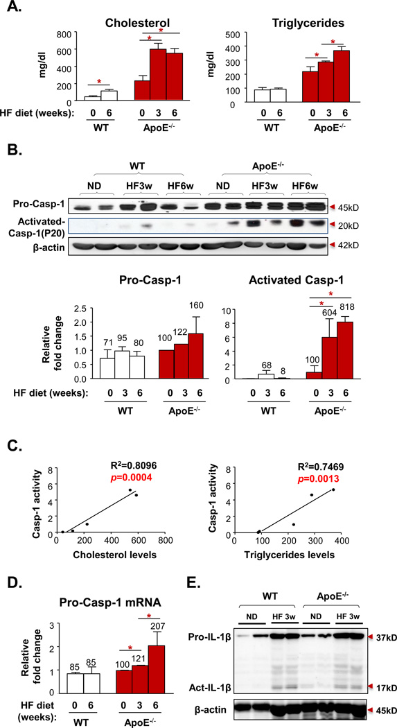
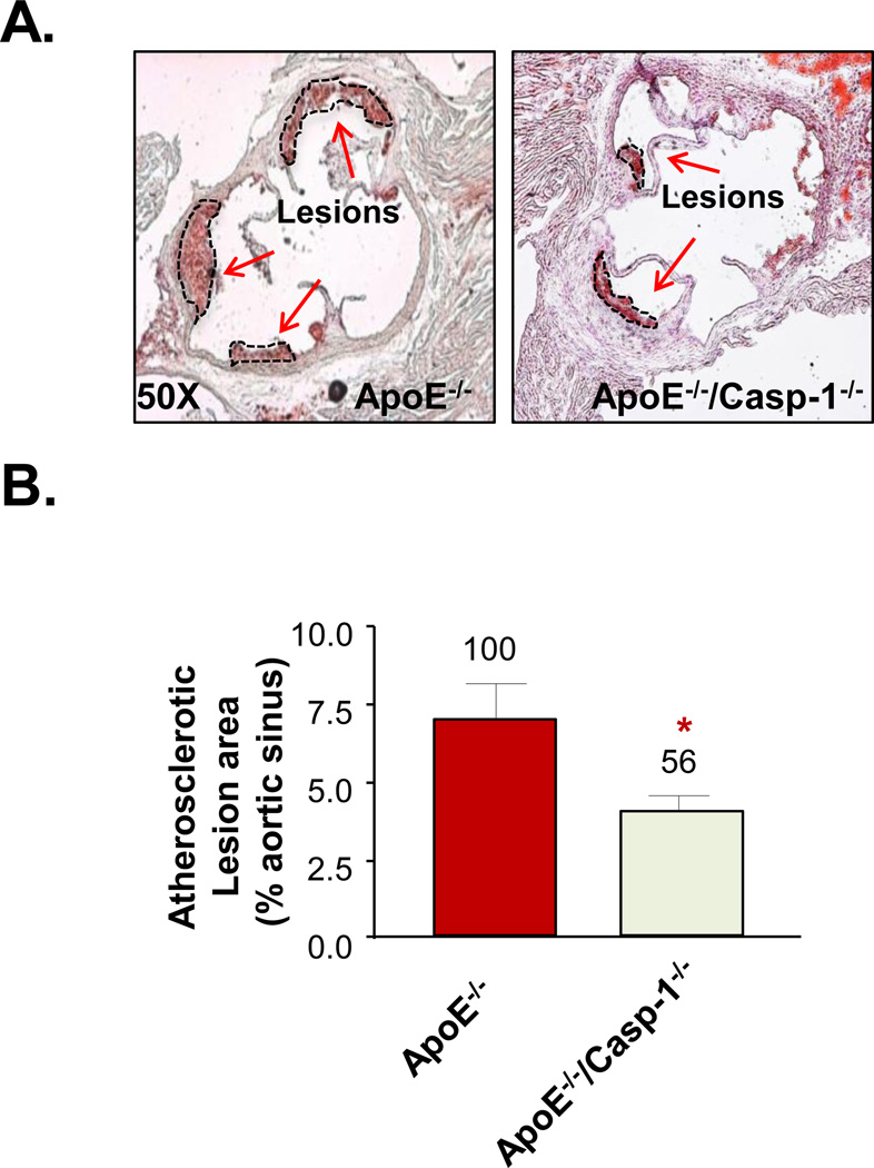
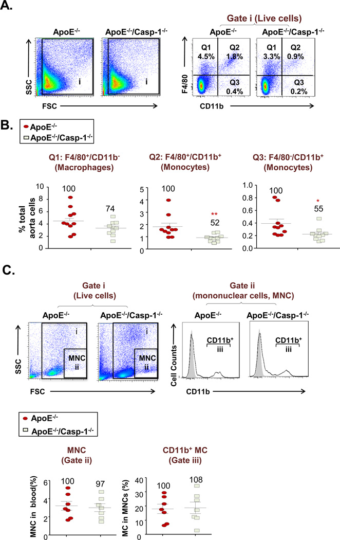
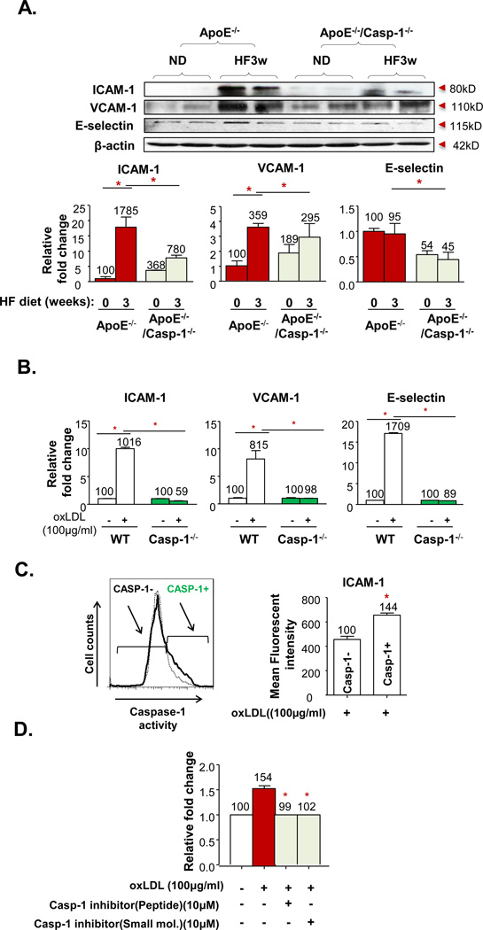
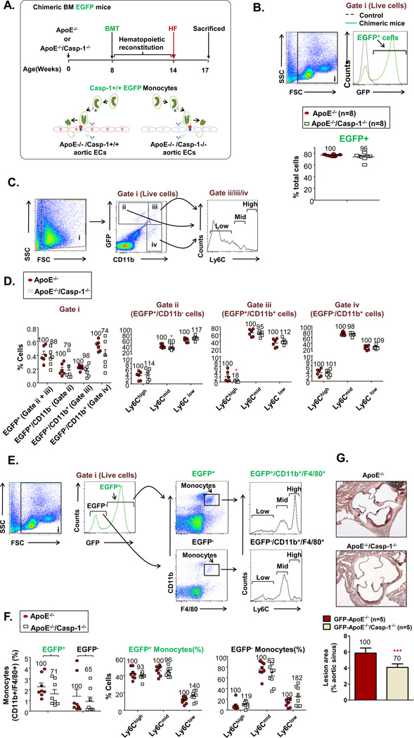
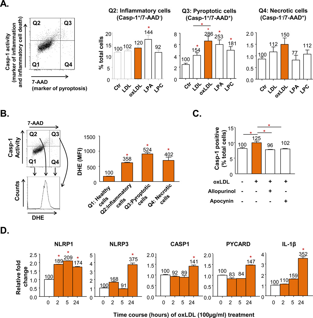
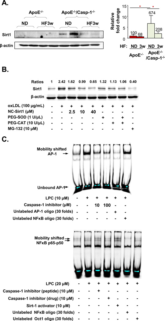
Approach and results: Using biochemical, immunologic, pathological, and bone marrow transplantation methods together with the generation of new apoplipoprotein E (ApoE)(-/-)/caspase-1(-/-) double knockout mice, we made the following observations: (1) early hyperlipidemia induced caspase-1 activation in ApoE(-/-) mouse aorta; (2) caspase-1(-/-)/ApoE(-/-) mice attenuated early atherosclerosis; (3) caspase-1(-/-)/ApoE(-/-) mice had decreased aortic expression of proinflammatory cytokines and attenuated aortic monocyte recruitment; and (4) caspase-1(-/-)/ApoE(-/-) mice had decreased EC activation, including reduced adhesion molecule expression and cytokine secretion. Mechanistically, oxidized lipids activated caspase-1 and promoted pyroptosis in ECs by a reactive oxygen species mechanism. Caspase-1 inhibition resulted in accumulation of sirtuin 1 in the ApoE(-/-) aorta, and sirtuin 1 inhibited caspase-1 upregulated genes via activator protein-1 pathway.

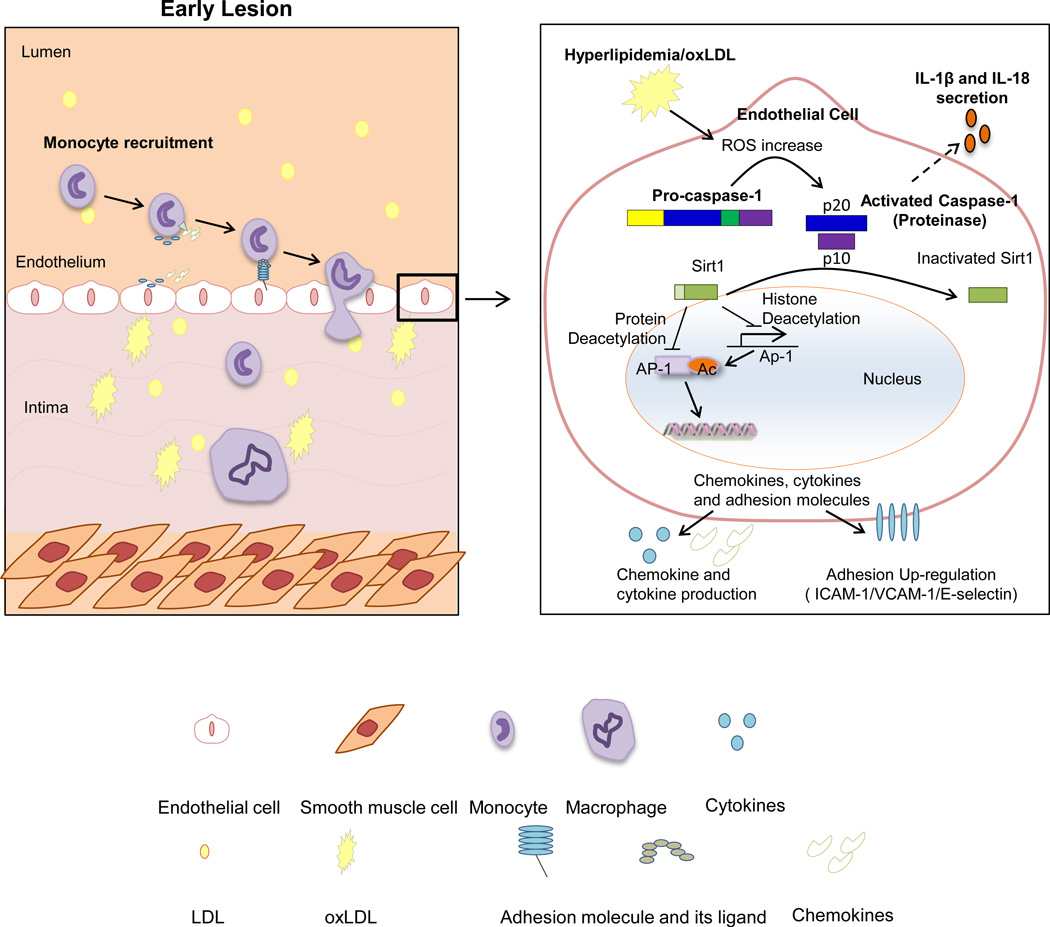
Conclusions: Our results demonstrate for the first time that early hyperlipidemia promotes EC activation before monocyte recruitment via a caspase-1-sirtuin 1-activator protein-1 pathway, which provides an important insight into the development of novel therapeutics for blocking caspase-1 activation as early intervention of metabolic cardiovascular diseases and inflammations.

Keywords: atherosclerosis; caspase-1; inflammation.


Figures

Figure 1
Figure 1. Early Hyperlipidemia Induces Caspase-1 (casp-1) Expression and Activation in Mouse Aorta
A. Plasma levels of cholesterol and triglycerides in wild type mice (WT) and apolipoprotein E gene deficient mice (ApoE−/−) after 0 week (ND), 3 weeks (HF3w), or 6 weeks (HF6w) of HF diet (n=5 for each group). B. The protein expression of pro-casp-1 and active casp-1 p20 subunit in mouse aorta lysate of WT and ApoE−/− mice after 0, 3, or 6 weeks of HF diet (n=2 for each group). C. Correlation of caps-1 activation and plasma lipid levels (of A and B). D. Casp-1 mRNA expression in aortas of WT and ApoE−/− after 0, 3, or 6 weeks of HF diet (n=3 for each group). E. The protein expression of pro-IL-1β and active IL-1β in mouse aorta lysate of WT and ApoE−/− mice with or without HF diet for 3 weeks. Data are expressed as mean ± SE. *, p<0.05, changes with the statistical significance.
Figure 2
Figure 2. Caspase-1 Deficiency Attenuates Early Atherosclerotic Lesion Formation in Aortic Sinus of ApoE−/−/Casp-1−/− Fed with a 3-week HF Diet
A. Representative images of atherosclerotic lesion staining of ApoE−/− (n=6) and ApoE−/−/Casp-1−/− mice (n=9) in mouse aortic sinus, as the arrows indicated. B. Atherosclerotic lesion quantification. Data are expressed as mean ± SE. *, p<0.05, changes with statistical significance.
Figure 3
Figure 3. Caspase-1 Deficiency Attenuates Monocyte Infiltration into Mouse Aorta in ApoE−/−/Casp-1−/− Mice fed with a 3-week HF Diet
A. Representative flow cytometric dot plots of live cells (Gate i) in mouse aortic single cell preparations. Monocytes were gated as CD11b+/F4/80+ and CD11b+/F4/80. Macrophages were gated as CD11b/F4/80+. B. Percentage of macrophages (CD11b/F4/80+), monocytes(F4/80+/CD11b+ and F4/80CD11b+) in total aortic cell population in ApoE−/− and ApoE−/−/Casp-1−/− mice after 3 weeks of a HF diet (n=10 for each group). C. Representative flow cytometric dot plots of live cells (Gate i) in mouse peripheral blood. Mononuclear cells (MNC, Gate ii) were first gated according the forward scatter (FSC) and side scatter (SSC). Monocytes (MC) were identified as CD11b+ mononuclear cells (Gate iii) (n=7 for each group). Data are expressed as mean ± SE. *, p<0.05, and **, p<0.01, indicate changes with the statistical significance.
Figure 4
Figure 4. Caspase-1 Activation Regulates Hyperlipidemia-Induced EC Activation in vivo and in vitro
A. Protein expression of ICAM-1, VCAM-1, and E-selectin in aortic tissues from ApoE−/− and ApoE−/−/Casp-1−/− mice after a HF diet for 3 weeks. Representative western blots (top). Quantification of protein expression normalized to the levels of β-actin (bottom). B. mRNA expressions of ICAM-1, VCAM-1, and E-selectin in mouse aortic endothelial cells (MAECs) from WT and Casp−/− mice, cultured and treated with oxLDL (100µg/mL) for 24 hours. C. Expression level of ICAM-1 in Casp-1 active human aortic endothelial cells (HAECs) after oxLDL treated for 6 hours. D. Effect of Casp-1 inhibition on oxLDL-induced monocytic THP-1 cell static adhesion to HAECs. HAECs were cultured and treated with oxLDL (100µg/mL) for 24 hours. Caspase-1 peptide inhibitor (10µM, z-YVAD-FMK) and caspase-1 small molecular inhibitor (10mM) were added 1 hr before the treatment. Data are expressed as mean ± SE. *, p<0.05.
Figure 5
Figure 5. Caspase-1 Deficient Aortas are Less Efficient in Recruiting Inflammatory Monocytes during Early Atherogenesis
A. Schematic representation of chimeric bone marrow (BM) EGFP mice generation. Casp-1+/+ BM cells collected from EGFP+ mice were injected into irradiated ApoE−/− mice or ApoE−/−/Casp-1−/− mice to determine the effect of caspase-1 deficiency in vascular cells on monocyte migration into the aorta. After a 6-week reconstitution period, the chimeric mice were fed with a HF diet for 3 weeks. B. The reconstitution rates of EGFP+ nuclear cells in the peripheral blood 6 weeks after BM transplantation. C. Monocyte population in mouse aorta after reconstitution with EGFP+ BM. Representative dot plots of CD11b/EGFP+ cells (Gate ii), CD11b+/EGFP+ monocytes (Gate iii), and CD11b+/EGFP (Gate iv) monocyte in mouse aorta. Monocytes in each of the three gates were further divided into 3 subsets: Ly-6Chigh, Ly-6Cmid, and Ly-6Clow. D. Quantification of EGFP+ and CD11b/EGFP+ cells, and CD11b+/EGFP+ and CD11b+/EGFP monocytes within live cells, and Ly-6Chigh, Ly-6Cmiddle, and Ly-6Clow monocytes within indicated gates in ApoE−/− and ApoE−/−/Casp-1−/− mouse aortas after BM reconstitution. Number within each graph represents cells in the ApoE−/−/Casp-1−/− mouse group as a percentage of the ApoE−/− mouse group (n=6 for each group). E. Monocyte population in mouse peripheral blood after reconstitution with EGFP+ BM. Representative dot plots of CD11b+/F4/80+ monocytes in both EGFP+ and EGFP peripheral blood cells. Monocytes were further divided into three subsets: Ly-6Chigh, Ly-6Cmid, and Ly-6Clow. F. Quantification of CD11b+/F4/80+ monocytes in both EGFP+ and EGFP peripheral blood cells and Ly-6Chigh, Ly-6Cmiddle, and Ly-6Clow cells in EGFP+ and EGFP monocytes. Number within each graph represents cells in the ApoE−/−/Casp-1−/− mouse group as a percentage of the ApoE−/− mouse group (n=8 for each group). G. Quantification of atherosclerotic lesion area in ApoE−/− and ApoE−/−/Casp-1−/− mouse aortas after BM reconstitution. Data are expressed as mean ± SE. *, p<0.05.
Figure 6
Figure 6. Oxidized Low-Density Lipoprotein (oxLDL) and its Components Induce Caspase-1 Activation in HAECs via a Reactive Oxygen Species (ROS)-Mediated Pathway
A. Pyroptotic cell death in HAECs caused by activation of casp1 induced by oxLDL and its components. HAECs were cultured and treated with LDL (100µg/mL), oxLDL(100µg/mL), oxLDL-derivatives lysophosphatidic acid (LPA, 100µM) or lysophosphatidylcholine (LPC, 15µM) as indicated for 6 hr. Casp-1 activity was determined by a commercial kit, and 7-Aminoactinomycin D (7-AAD) fluorescence dye was used to determine the cell membrane integrity. casp-1+/7-AAD+ cells were gated as pyrototic cells (Q3), casp-1+ single positive cells (Q2) were gated as inflammatory cells, and 7-AAD+ single positive cells (Q4) were gated as necrotic cells. B. ROS levels in pyroptotic cells. ROS levels were determined by dihydroethidium (DHE) fluorescence dye staining and the mean fluorescence intensity (MFI) of DHE+ cell fraction was determined. C. Attenuation of oxLDL-induced caspase-1 activation in HAECs with ROS inhibitors Allopurinol (xanthine oxidase inhibitor) and Apocynin (NADPH oxidase inhibitor). Allopurinol (1mM) and Apocynin (100µM) were added 1 hour before oxLDL treatment. HAECs were then treated with oxLDL (100µg/mL) for 6 hr and stained for caspase-1 activity. D. mRNA upregulation of inflammasome components including NLRP1 (Nod-like receptor protein 1), NLRP3 (Nod-like receptor 3), PYCARD (or ASC, inflammasome adaptor Apoptosis-associated speck-like protein containing a CARD), caspase-1, and IL-1β (interleukin-1β) in HAECs treated with oxLDL. Data are expressed mean ± SE. *; p<0.05, changes with statistical significance.
Figure 7
Figure 7. Hyperlipidemia/dyslipidemia Decreases Sirtuin 1 (Sirt1) Expression in ApoE−/− Mouse Aorta and Induces Sirt1 Cleavage in Human Aortic ECs through Caspase-1 Activation
A. Caspase-1 deficiency results in Sirt1 accumulation in mouse aorta in ApoE−/−/Casp-1−/− mice. ApoE−/− mice and ApoE−/−/Casp-1−/− mice were fed with a HF diet for 3 weeks. Mouse aortic tissues were collected for uncleaved Sirt1 protein expression analysis by Western blot with the specific antibody (left panel). The quantification of Sirt1 expression in the Western blot was presented after normalized with the expression of β-actin in the same sample (right panel). B. OxLDL induces Sirt1 cleavage in HAECs. HAECs were cultured and treated with oxLDL (100µg/mL) for 24 hr. Different doses of non-caspase-1 cleavable sirt1 peptide (NC-SIRT1), superoxide scavengers PEG-SOD and PEG-CAT, and proteasome inhibitor MG-132 were added 1 hr before oxLDL treatment. The protein lysates were collected, and cleaved-Sirt1 expression was determined by Western blot with anti-Sirt1 antibody. The relative changes of Sirt1 expression normalized by β-actin (ratios) were calculated based on the ratios of Sirt1 expression levels in treated samples over that in non-treated samples. C. Inhibition of caspase-1 attenuates LPC induced AP-1 binding to the AP-1 site, revealed by AP-1 electrophoretic gel mobility shift assay (upper panel), whereas inhibition of caspase-1 does not decrease NF-kB binding to the NF-kB site (lower panel). Data are expressed mean ± SE. *; p<0.05, changes with statistical significance.
Figure 8
Figure 8. Schematic representation of a new model for the role of caspase-1 activation in ECs during early atherogenesis

Similar articles

Cited by

References

    1. Libby P, Ridker PM, Hansson GK. Progress and challenges in translating the biology of atherosclerosis. Nature. 2011;473:317–325. - PubMed
    1. Mestas J, Ley K. Monocyte-endothelial cell interactions in the development of atherosclerosis. Trends in cardiovascular medicine. 2008;18:228–232. - PMC - PubMed
    1. Jiang X, Yang F, Tan H, Liao D, Bryan RM, Jr, Randhawa JK, Rumbaut RE, Durante W, Schafer AI, Yang X, Wang H. Hyperhomocystinemia impairs endothelial function and enos activity via pkc activation. Arterioscler Thromb Vasc Biol. 2005;25:2515–2521. - PMC - PubMed
    1. Zhang D, Jiang X, Fang P, Yan Y, Song J, Gupta S, Schafer AI, Durante W, Kruger WD, Yang X, Wang H. Hyperhomocysteinemia promotes inflammatory monocyte generation and accelerates atherosclerosis in transgenic cystathionine beta-synthase-deficient mice. Circulation. 2009;120:1893–1902. - PMC - PubMed
    1. Combadiere C, Potteaux S, Rodero M, Simon T, Pezard A, Esposito B, Merval R, Proudfoot A, Tedgui A, Mallat Z. Combined inhibition of ccl2, cx3cr1, and ccr5 abrogates ly6c(hi) and ly6c(lo) monocytosis and almost abolishes atherosclerosis in hypercholesterolemic mice. Circulation. 2008;117:1649–1657. - PubMed

Publication types

MeSH terms