. 2015 Feb 23;10(2):e0118311.
doi: 10.1371/journal.pone.0118311. eCollection 2015.

Expression of the SNARE protein SNAP-23 is essential for cell survival

Affiliations

Expression of the SNARE protein SNAP-23 is essential for cell survival

Sunil Kaul et al. PLoS One. .

Abstract

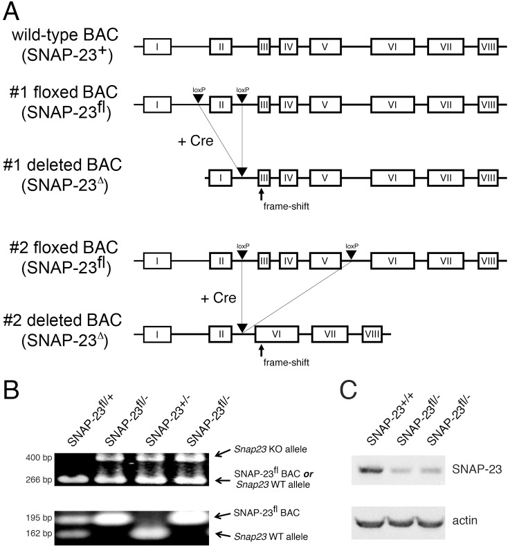
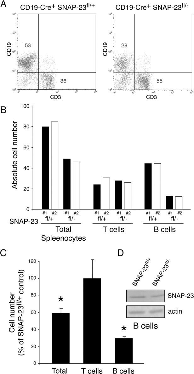
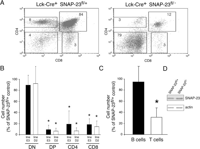
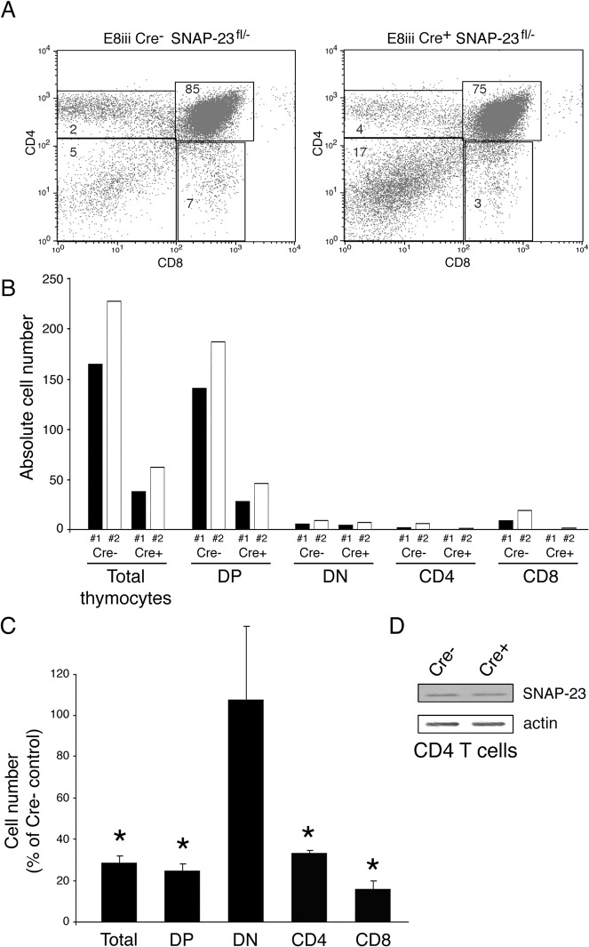
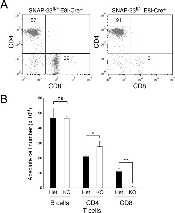
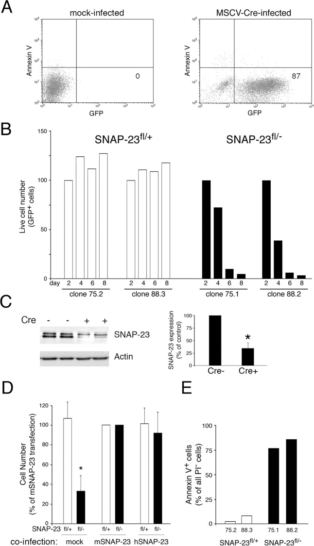
Members of the SNARE-family of proteins are known to be key regulators of the membrane-membrane fusion events required for intracellular membrane traffic. The ubiquitously expressed SNARE protein SNAP-23 regulates a wide variety of exocytosis events and is essential for mouse development. Germline deletion of SNAP-23 results in early embryonic lethality in mice, and for this reason we now describe mice and cell lines in which SNAP-23 can be conditionally-deleted using Cre-lox technology. Deletion of SNAP-23 in CD19-Cre expressing mice prevents B lymphocyte development and deletion of SNAP-23 using a variety of T lymphocyte-specific Cre mice prevents T lymphocyte development. Acute depletion of SNAP-23 in mouse fibroblasts leads to rapid apoptotic cell death. These data highlight the importance of SNAP-23 for cell survival and describe a mouse in which specific cell types can be eliminated by expression of tissue-specific Cre-recombinase.


Conflict of interest statement

Competing Interests: The authors have declared that no competing interests exist.

Figures

Fig 1
Fig 1. Generation of mice containing a floxed SNAP-23 allele.
(A) A 179 kb BAC clone containing the entire SNAP-23 gene was mutated by introducing loxP sites surrounding exon II alone (#1 floxed allele) or surrounding exons III-V (#2 floxed allele). Expression of Cre will result in the excision of exon II alone in the #1 floxed allele construct or exons III-V in #2 floxed allele construct. Each deleted allele introduces a frame shift that prevents the synthesis of in-frame SNAP-23 exon-encoded amino acids. (B) Typical PCR genotyping analysis of mice used in this study. The upper panel shows results obtained using a PCR reaction that can distinguish the deleted SNAP-23 allele present in SNAP-23+/- mice (492 bp) from either the wild-type SNAP-23 allele or the floxed SNAP-23 BAC transgene (266 bp). The lower panel shows results obtained using a PCR reaction that can distinguish the floxed SNAP-23 BAC transgene (195 bp) from the wild-type SNAP-23 allele present in SNAP-23+/- mice (161 bp). (C) Spleen cells were isolated from wild-type mice (SNAP-23+/+) and two representative floxed SNAP-23 BAC transgene mice on a SNAP-23-/- background (SNAP-23fl/-) and the cells were lysed and analyzed by SDS-PAGE and immunobloting with SNAP-23 and actin antibodies. Quantitative densitometry revealed that the floxed SNAP-23 BAC expression of SNAP-23 was approximately 50% that found in wild-type mice.
Fig 2
Fig 2. SNAP-23 is essential for B cell survival.
Spleens were harvested from CD19-Cre+ SNAP-23fl/+ control mice or CD19-Cre+ SNAP-23fl/- mice (line E3) and single cell suspensions were stained with fluorochrome-conjugated CD19- and CD3-mAb and analyzed by flow cytometry. (A) A representative flow cytometry profile reveals a dramatic reduction in the percentage of CD19+ B cells in the spleen. (B) Quantitative analysis of two different matched pairs of CD19-Cre+ SNAP-23fl/+ or SNAP-23fl/- littermate mice indicating total spleen cell numbers as well as the numbers of T cells (CD3+ cells) and B cells (CD19+ cells) present in each spleen (calculated based on the percentage of each cell type present as determined by flow cytometry). (C) Summary of three independent experiments showing the recovery of the indicated cell types from the spleens of CD19-Cre+ SNAP-23fl/- mice expressed as a percentage of those found in CD19-Cre+ SNAP-23fl/+ control mouse spleens. The data shown are mean +/- SD of three independent experiments (*p<0.05). (D) B cells were purified from the spleens from the indicated mice and equal numbers of cell equivalents were analyzed by SDS-PAGE and immunoblotting using a SNAP-23 antibody. The blot was re-probed for anti-β actin mAb as a loading control.
Fig 3
Fig 3. Deletion of SNAP-23 in Lck-expressing T cells prevents T cell development.
Thymi were harvested from Lck-Cre+ SNAP-23fl/+ control mice or Lck-Cre+ SNAP-23fl/- mice and single cell suspensions were stained with fluorochrome-conjugated CD4- and CD8-mAb and analyzed by flow cytometry. (A) A representative flow cytometry profile reveals a dramatic reduction in the percentage of CD4+, CD4+8+, and CD8+ T cells in the thymus of an Lck-Cre+-expressing SNAP-23fl/- mouse (line D2). (B) Quantitative analysis of matched pairs of littermate mice from SNAP-23fl founder lines E3 or D2 indicate recovery of CD4-CD8- (DN), CD4+CD8+ (DP), CD4+, and CD8+ T cells present in thymi of Lck-Cre+ SNAP-23fl/- mice expressed as a percentage of those found in thymi of Lck-Cre+ SNAP-23fl/+ control mice (calculated based on the percentage of each cell type present as determined by flow cytometry). The data shown are mean +/- SD of three independent experiments (*p<0.05). (C) The recovery of the indicated cell types from the spleens of Lck-Cre+ SNAP-23fl/- mice was expressed as a percentage of those found in Lck-Cre+ SNAP-23fl/+ control mouse spleens (line D5). The data shown are mean +/- SD of eight independent experiments (*p<0.05). (D) T cells were purified from the spleens from the indicated mice and equal numbers of cell equivalents were analyzed by SDS-PAGE and immunoblotting using a SNAP-23 antibody. The blot was re-probed for anti-β actin mAb as a loading control.
Fig 4
Fig 4. Deletion of SNAP-23 in CD4+CD8+ T cells prevents T cell development.
Thymi were harvested from E8iii-Cre- SNAP-23fl/- control mice or E8iii-Cre+ SNAP-23fl/- mice (line E3) and single cell suspensions were stained with fluorochrome-conjugated CD4- and CD8-mAb and analyzed by flow cytometry. (A) A representative flow cytometry profile reveals a dramatic reduction in the percentage of CD4+, CD4+8+, and CD8+ T cells in the thymus of an E8iii-Cre+-expressing SNAP-23fl/- mouse. (B) Quantitative analysis of matched pairs of Cre- and Cre+ SNAP-23fl/- littermate mice lines indicate recovery of CD4-CD8- (DN), CD4+CD8+ (DP), CD4+, and CD8+ T cells present in thymi of E8iii-Cre+ SNAP-23fl/- mice expressed as a percentage of those found in thymi of E8iii-Cre- SNAP-23fl/- control mice (calculated based on the percentage of each cell type present as determined by flow cytometry). (C) The recovery of the indicated cell types from the thymi of E8iii-Cre+ SNAP-23fl/- mice was expressed as a percentage of those found in E8iii-Cre- SNAP-23fl/- control mouse spleens. The data shown are mean +/- SD of five independent experiments (*p<0.05). (D) CD4 T cells were purified from the thymi from the indicated mice and equal numbers of cell equivalents were analyzed by SDS-PAGE and immunoblotting using a SNAP-23 antibody. The blot was re-probed for anti-β actin mAb as a loading control.
Fig 5
Fig 5. Deletion of SNAP-23 in thymus-derived CD8 T cells prevents survival of mature CD8 T cells in the spleen.
Spleens were harvested from E8i-Cre+ SNAP-23fl/+ control mice or E8i-Cre+ SNAP-23fl/- mice (line E3) and single cell suspensions were stained with fluorochrome-conjugated CD3-, CD4-, and CD8-mAb and analyzed by flow cytometry. (A) A representative flow cytometry profile of CD3-gated spleen cells reveals a selective reduction in the percentage of CD8+ T cells in the spleen. (B) Quantitative analysis of E8i-Cre+ SNAP-23fl/+ or E8i-Cre+ SNAP-23fl/- littermate mice indicating total spleen cell numbers, B cell numbers (determined by gating on B220+ spleen cells) as well as the numbers of CD4 T cells and CD8 T cells present in each spleen (calculated based on the percentage of each cell type present as determined by flow cytometry). The data shown are mean +/- SD of three independent experiments (*p<0.05; **, p<0.01).
Fig 6
Fig 6. Deletion of SNAP-23 leads to acute death of MEFs.
MEF lines were generated from SNAP-23fl/+ mice (clones 75.1 and 88.2) or SNAP-23fl/- mice (clones 75.2 and 88.3). (A) Infection of a typical SNAP-23fl/+ MEF culture infected with empty retrovirus (left panel) or GFP-Cre retrovirus (right panel) shows that two days after infection the vast majority of MEFs were viable (Annexin V-) and expressed GFP-Cre. (B) The indicated MEF lines were infected with GFP-Cre-expressing retrovirus and the number of live cells present in each culture (based on staining with PI) was determined at different times. The absolute cell recovery in each condition was expressed relative to the amount of cells present two days after infection (control experiments showed that there was no Cre-dependent cell death in any line after only two days of infection). The data shown are representative of two independent experiments analyzed at day 2, 4, 6, 8 and one experiment analyzed at day 1, 3, 5, 7. (C) Adherent SNAP-23fl/- MEFs were isolated 4 days after retroviral transduction with Cre (Cre+) or after mock-transduction (Cre-). Equal numbers of cells from each culture were analyzed by SDS-PAGE and immunoblotting using a SNAP-23 antibody. The blot was re-probed for anti-β actin mAb as a loading control. The amount of SNAP-23 present in each cell lysate was normalized to the amount of actin present and the data shown are mean +/- SD of three independent experiments (*p<0.05). (D) MEF lines generated from SNAP-23fl/+ mice or SNAP-23fl/- mice were co-infected with retrovirus containing GFP-Cre and retrovirus containing nothing (mock), mouse SNAP-23 (mSNAP-23), or human SNAP-23 (hSNAP-23). After 4 days the number of live cells present in each culture (based on staining with PI) was determined and was expressed relative to the number of cells recovered in the mSNAP-23 co-infection condition. The data shown are mean +/- SD of four independent experiments (*p<0.05). (E) The indicated MEF lines were infected with GFP-Cre-expressing retrovirus and after four days the cells were stained with PI and Annexin V and analyzed for GFP-Cre expression. The percentage of viable (PI-) GFP-Cre+ Annexin V+ cells in each culture was determined by flow cytometry. The data shown are representative of three independent experiments.

References

    1. Jahn R, Scheller RH. SNAREs—engines for membrane fusion. Nat Rev Mol Cell Biol. 2006; 7: 631–643. - PubMed
    1. Washbourne P, Thompson PM, Carta M, Costa ET, Mathews JR, Lopez-Bendito G, et al. Genetic ablation of the t-SNARE SNAP-25 distinguishes mechanisms of neuroexocytosis. Nat Neurosci. 2002; 5: 19–26. - PubMed
    1. Suh YH, Terashima A, Petralia RS, Wenthold RJ, Isaac JT, Roche KW, et al. A neuronal role for SNAP-23 in postsynaptic glutamate receptor trafficking. Nat Neurosci. 2010; 13: 338–343. 10.1038/nn.2488 - DOI - PMC - PubMed
    1. Foster LJ, Yaworsky K, Trimble WS, Klip A. SNAP23 promotes insulin-dependent glucose uptake in 3T3-L1 adipocytes: possible interaction with cytoskeleton. Am J Physiol. 1999; 276: C1108–1114. - PubMed
    1. Guo Z, Turner C, Castle D. Relocation of the t-SNARE SNAP-23 from lamellipodia-like cell surface projections regulates compound exocytosis in mast cells. Cell. 1998; 94: 537–548. - PubMed

Publication types

MeSH terms

LinkOut - more resources