. 2015 Feb 23;10(2):e0117518.
doi: 10.1371/journal.pone.0117518. eCollection 2015.

SATB1 overexpression regulates the development and progression in bladder cancer through EMT

Affiliations

SATB1 overexpression regulates the development and progression in bladder cancer through EMT

Feng Wan et al. PLoS One. .

Retraction in

Abstract

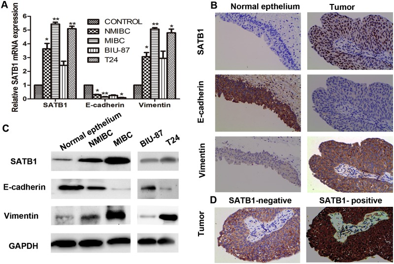
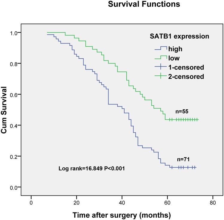
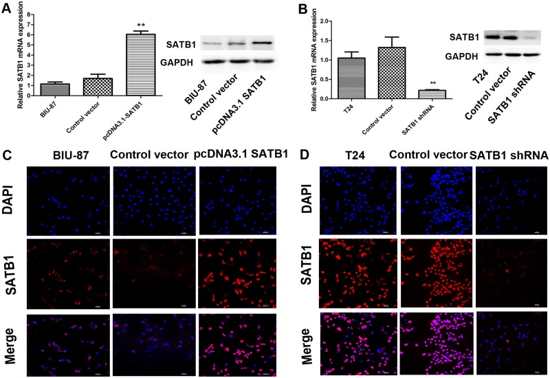
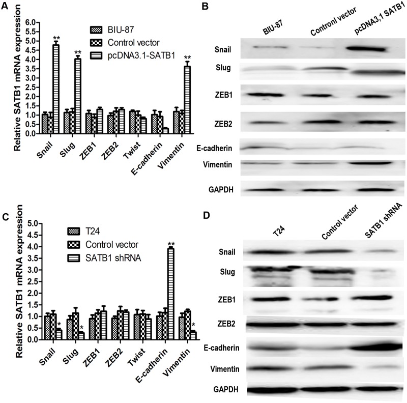
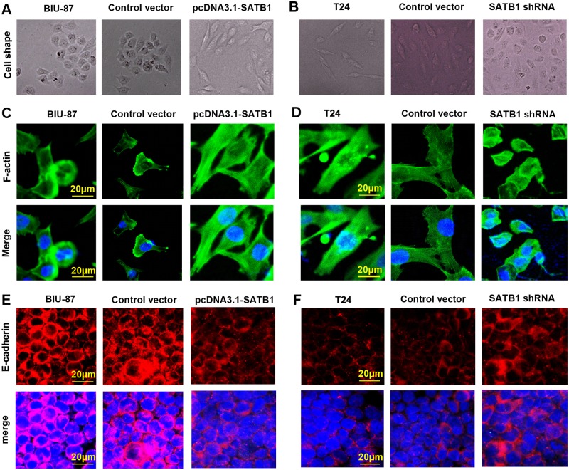
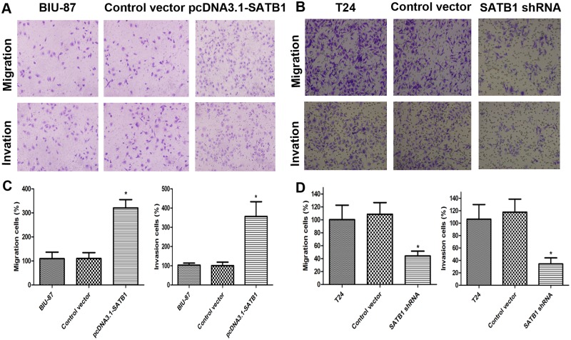
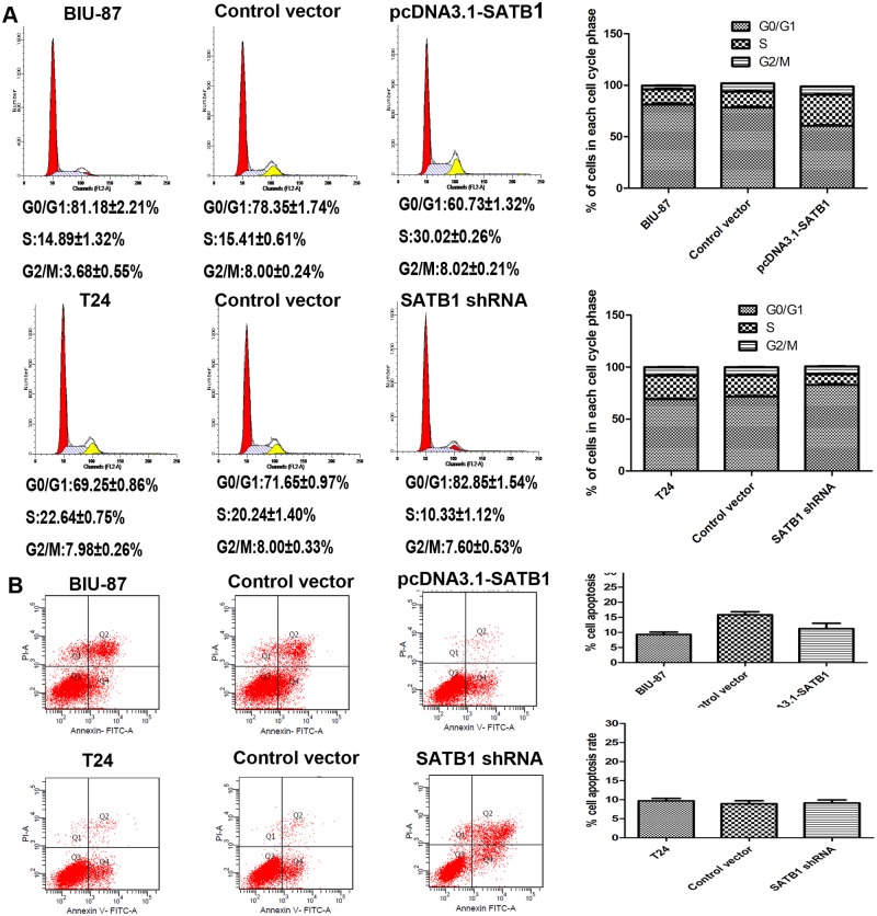
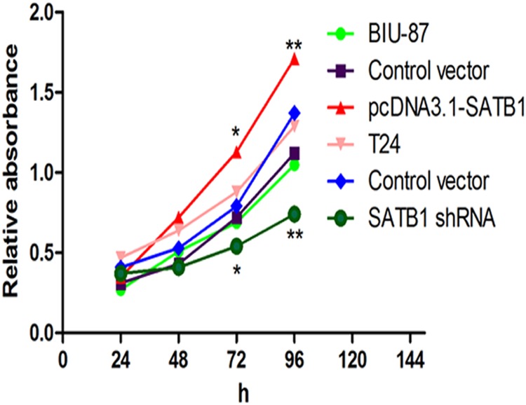
The global gene regulator Special AT-rich sequence-binding protein-1 (SATB1) has been reported to induce EMT-like changes and be associated with poor clinical outcome in several cancers. This study aims to evaluate whether SATB1 affects the biological behaviors of bladder transitional cell carcinoma (BTCC) and further elucidate if this effect works through an epithelial-mesenchymal transition (EMT) pathway. The expression of SATB1, E-cadherin (epithelial markers), vimentin (mesenchymal markers) in BTCC tissues and adjacent noncancerous tissues, as well as in two cell lines of bladder cancer were investigated. Whether the SATB1 expression is associated with clinicopathological factors or not was statistically analyzed. Cell invasion and migration, cell cycle, cell proliferation and apoptosis were evaluated in SATB1 knockdown and overexpressed cell lines. Our results showed that the expression of SATB1 was remarkably up-regulated both in BTCC tissues and in bladder cancer cell lines with high potential of metastasis. The results were also associated with EMT markers and poor prognosis of BTCC patients. Moreover, SATB1 induced EMT processes through downregulation of E-cadherin, upregulation of E-cadherin repressors (Snail, Slug and vimentin). SATB1 also promoted cell cycle progression, cell proliferation, cell invasion and cell migration, but did not alter cell survival. In conclusion, our results suggest that SATB1 plays a crucial role in the progression of bladder cancer by regulating genes controlling EMT processes. Further, it may be a novel therapeutic target for aggressive bladder cancers.


Conflict of interest statement

Competing Interests: The authors have declared that no competing interests exist.

Figures

Fig 1
Fig 1. SATB1 is upregulated in human bladder carcinoma tissues and bladder carcinoma cell lines with high metastatic potential.
Expressions of mRNA levels of SATB1, E-cadherin and vimentin in human bladder carcinoma tissues and two bladder carcinoma cell lines were assessed by quantitative RT-PCR (A; *P < 0.05, **P < 0.001). IHC staining of human bladder carcinoma tissues and corresponding adjacent non-cancerous tissues for SATB1, E-cadherin and vimentin was performed. SATB1 staining was observed in the nuclei of bladder cancer cells, vimentin was found to be predominantly stained in plasmalemma of bladder cancer cells, but E-cadherin staining was observed mainly in plasmalemma of non-cancerous bladder cells (B); Bladder cancer samples with SATB1-positive were stained with vimentin but not E-cadherin. However, tumor samples with SATB1-negative were stained with E-cadherin but not vimentin (D). The original magnification was ×400x (B and D). Protein levels of SATB1, E-cadherin and vimentin were examined by western blot (C). Error bars indicate s.e.m., n = 3 experiments.
Fig 2
Fig 2. Kaplan-Meier estimates of survival according to SATB1 expression in patients with bladder cancer.
The 5-year overall survival rate in patients with high SATB1 expression was significantly worse than those with low SATB1 expression (P<0.001; log-rank test).
Fig 3
Fig 3. The SATB1-overexpressing cells and SATB1-knockdown cells are established.
The SATB1 expression of BIU-87 cells and T24 cells transfected with pcDNA3.1-SATB1 and pGenesil2-SATB1-shRNA were examined by qRT-PCR and western blot (A and B; **P < 0.001). Non-transfected BIU-87 cells were used as control group. T24 cells treated with pGenesil2 control vector which does not target any specific gene were used as the control groups. Immunofluorescence analysis was performed to detect the SATB1staining in each cell groups. Immunofluorescence images at 200×magnification (C and D). Error bars indicate s.e.m., n = 3 experiments.
Fig 4
Fig 4. Ectopic expression of SABT1 induces EMT and knockdown of SATB1 reverses EMT in established human cancer cell lines.
The expression of Snail and Slug, two inhibitors of E-cadherin, and the mesenchymal marker vimentin were upregulated after SATB1 expression was enhanced (A; **P<0.001). The mRNA levels of E-cadherin expression were decreased after transfection of pcDNA3.1-SATB1 expression plasmids (A; P < 0.05). There was a trend for reduction in E-cadherin protein expression in BIU-87 cells with enhanced SATB1 expression (B; P>0.05). After SATB1 expression was silenced with SATB1 shRNA in T24 cells, the expression of Snail, Slug and mesenchymal markers vimentin was downregulated. The expression of epithelial marker, E-cadherin, was upregulated compared to nontransfected cells and control vector-transfected cells (C and D; *P<0.05;**P<0.001). There were no remarkable changes in the expressions of ZEB1, ZEB2 and Twist, other known inhibitors of E-cadherin, in BIU-87 with enhanced SATB1 expression and T24 cells with reduced SATB1 expression, respectively.
Fig 5
Fig 5. SATB1 induces EMT-like changes in bladder cancer cells.
BIU-87 cells with enhanced SATB1 expression exhibited a marked change, from a cobblestone-shaped morphology to a spindle-shaped morphology (A and C), a classical marker of EMT. T24 cells with reduced SATB1 expression showed a major cell morphological change, from a spindle-like fibroblastic morphology to a cobble-stone-like morphology (B and D), consistent with the cells undergoing MET. The images of cell morphological changes were collected using an inverted microscope at 200×magnification(A and B) and A1Si laser-scanning confocal microscope at 400×magnification (C and D; scale bar = 20 μm) in each group cells. Photographs of representative cells are shown (C and D). The E-cadherin was reduced on the plasma membrane in the established pcDAN3.1-SATB1 BIU-87 cells, but was founded to be enriched in SATB1-shRNA T24 cells (E and F).
Fig 6
Fig 6. Cell invasion and migration capability in vitro were detected by transwell invasion and migration assay.
The cells invading the lower chamber were stained with crystal violet, photographed, and then extracted with 10% acetic acid. Representative images of cell migrations to the bottom chamber are shown (A and B). BIU-87 cells with enhanced SATB1 expression exhibited increased invasiveness and T24 cells with reduced SATB1 expression showed reduced invasiveness, when compared to control groups (C and D;*P < 0.05). Error bars indicate s.e.m., n = 3 experiments.
Fig 7
Fig 7. Effects of SATB1 on cell cycle progression and apoptosis of BIU-87 and T24 cells in vitro.
The cell population at each stage of the cell cycle and apoptosis were analyzed by flow cytometry. After SATB1 expression was enhanced with pcDNA3.1-SATB1 plasmid in BIU-87 cells, the ratio of cells in G0/G1 phase decreased, and the ratio of cells entering S phase increased significantly compared to nontransfected cells and control cells (A; *P < 0.05). After SATB1 expression was silenced with SATB1 shRNA in T24 cells, the cell cycle progression was arrested in G0/G1 phase, and the ratio of cells entering S phase was reduced significantly compared to nontransfected cells and control cells (A; *P < 0.05).There were no changes in the apoptosis rate in SATB1-overexpressing BIU-87 cells and in SATB1-lowexpressing T24 cells (B; P > 0.05).
Fig 8
Fig 8. Cell proliferation rates were measured by CCK-8 cell proliferation assay.
Absorbance at 450 nm was determined at the indicated time points and 630 nm was the reference wavelength (*P < 0.05, **P < 0.001), Error bars indicate s.e.m., n = 3 experiments.

References

    1. Jemal A, Bray F, Center MM, Ferlay J, Ward E, et al. (2011) Global cancer statistics. CA Cancer J Clin 61: 69–90. 10.3322/caac.20107 - DOI - PubMed
    1. Yang GL, Zhang LH, Bo JJ, Huo XJ, Chen HG, et al. (2012) Increased expression of HMGB1 is associated with poor prognosis in human bladder cancer. J Surg Oncol 106: 57–61. 10.1002/jso.23040 - DOI - PubMed
    1. Lei Y, Yan S, Ming-De L, Na L, Rui-Fa H (2011) Prognostic significance of Aurora-A expression in human bladder cancer. Acta Histochem 113: 514–518. 10.1016/j.acthis.2010.05.004 - DOI - PubMed
    1. Naito S, Bilim V, Yuuki K, Ugolkov A, Motoyama T, et al. (2010) Glycogen synthase kinase-3beta: a prognostic marker and a potential therapeutic target in human bladder cancer. Clin Cancer Res 16: 5124–5132. 10.1158/1078-0432.CCR-10-0275 - DOI - PubMed
    1. Wu CF, Ng KF, Chen CS, Chang PL, Chuang CK, et al. (2010) Expression of parvin-beta is a prognostic factor for patients with urothelial cell carcinoma of the upper urinary tract. Br J Cancer 103: 852–860. 10.1038/sj.bjc.6605835 - DOI - PMC - PubMed

Publication types

Substances