. 2015 Feb 23;10(2):e0117447.
doi: 10.1371/journal.pone.0117447. eCollection 2015.

Granulocyte-macrophage colony stimulatory factor enhances the pro-inflammatory response of interferon-γ-treated macrophages to Pseudomonas aeruginosa infection

Affiliations

Granulocyte-macrophage colony stimulatory factor enhances the pro-inflammatory response of interferon-γ-treated macrophages to Pseudomonas aeruginosa infection

Sonali Singh et al. PLoS One. .

Abstract

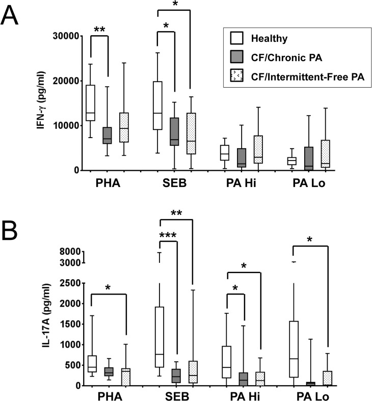
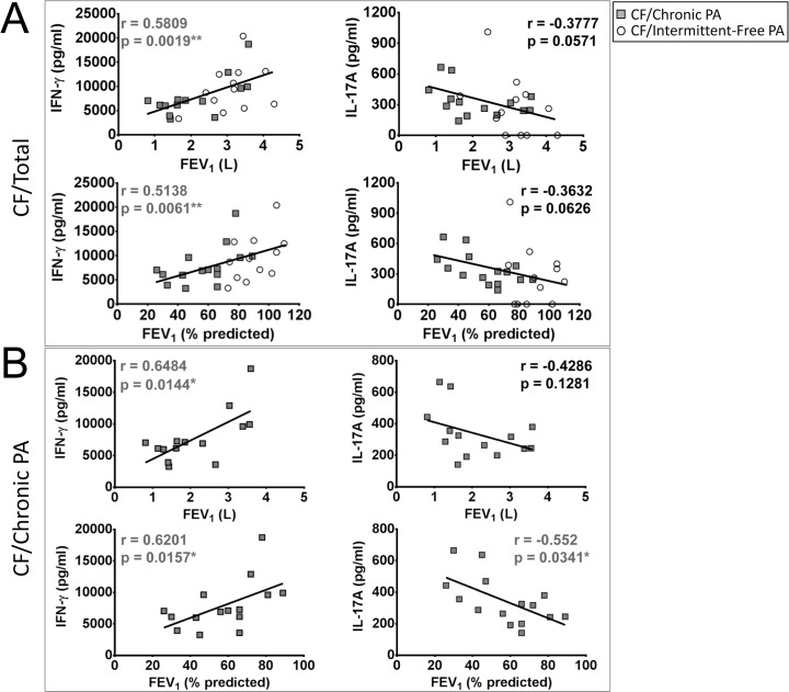
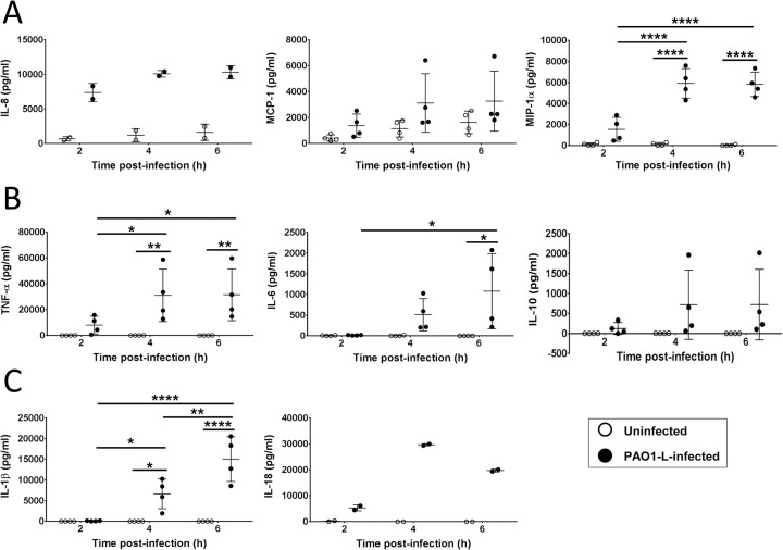
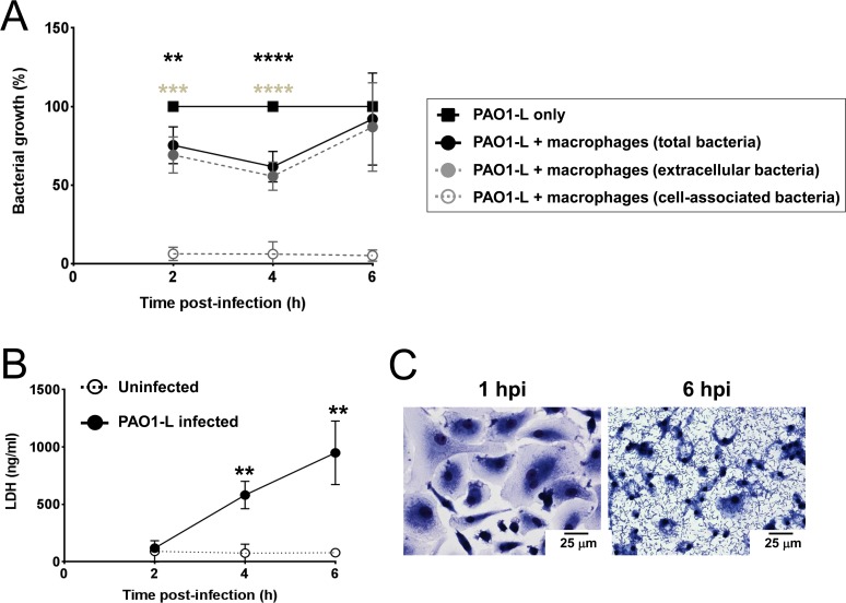
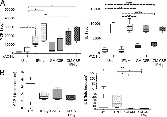
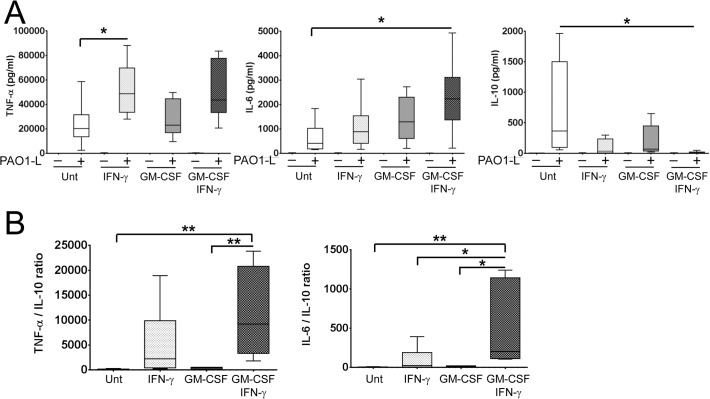
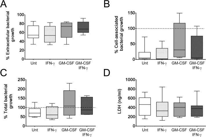
Pseudomonas aeruginosa is an opportunistic pathogen that can cause severe infections at compromised epithelial surfaces, such those found in burns, wounds, and in lungs damaged by mechanical ventilation or recurrent infections, particularly in cystic fibrosis (CF) patients. CF patients have been proposed to have a Th2 and Th17-biased immune response suggesting that the lack of Th1 and/or over exuberant Th17 responses could contribute to the establishment of chronic P. aeruginosa infection and deterioration of lung function. Accordingly, we have observed that interferon (IFN)-γ production by peripheral blood mononuclear cells from CF patients positively correlated with lung function, particularly in patients chronically infected with P. aeruginosa. In contrast, IL-17A levels tended to correlate negatively with lung function with this trend becoming significant in patients chronically infected with P. aeruginosa. These results are in agreement with IFN-γ and IL-17A playing protective and detrimental roles, respectively, in CF. In order to explore the protective effect of IFN-γ in CF, the effect of IFN-γ alone or in combination with granulocyte-macrophage colony-stimulating factor (GM-CSF), on the ability of human macrophages to control P. aeruginosa growth, resist the cytotoxicity induced by this bacterium or promote inflammation was investigated. Treatment of macrophages with IFN-γ, in the presence and absence of GM-CSF, failed to alter bacterial growth or macrophage survival upon P. aeruginosa infection, but changed the inflammatory potential of macrophages. IFN-γ caused up-regulation of monocyte chemoattractant protein-1 (MCP-1) and TNF-α and down-regulation of IL-10 expression by infected macrophages. GM-CSF in combination with IFN-γ promoted IL-6 production and further reduction of IL-10 synthesis. Comparison of TNF-α vs. IL-10 and IL-6 vs. IL-10 ratios revealed the following hierarchy in regard to the pro-inflammatory potential of human macrophages infected with P. aeruginosa: untreated < treated with GM-CSF < treated with IFN-γ < treated with GM-CSF and IFN-γ.


Conflict of interest statement

Competing Interests: The authors have declared that no competing interests exist.

Figures

Fig 1
Fig 1. Production of IFN-γ and IL-17A in CF and control PBMCs in response to several stimuli.
PBMCs were stimulated with 2% (v/v) phytohaemagglutinin (PHA), 10 ng/ml staphylococcal enterotoxin B (SEB), or two concentrations of PAO1-L lysates—PA Hi (6,250 ng/ml total protein) and PA Lo (24 ng/ml total protein)—for 6 days, re-stimulated with 15 ng/ml phorbol 12-myristate 13-acetate (PMA) overnight, and supernatants collected on day 7 for cytokine analysis. A. CF patient PBMCs produced less IFN-γ than healthy control PBMCs in response to PHA and SEB, but not PAO1-L lysates. B. CF patient PBMCs produced less IL-17A than healthy control PBMCs in response to PHA, SEB and PAO1-L lysates. Graph depicts 5–95 percentile with median; healthy controls: n = 13, CF/Chronic PA and CF/Intermittent-Free PA: n = 15 each. Significance calculated by Kruskal-Wallis test with Dunn’s post test; * = p≤0.05, ** = p≤0.01, *** = p≤0.001.
Fig 2
Fig 2. Correlation between IFN-γ and IL-17A production by PHA-stimulated PBMC cultures and lung function in CF patients.
A. Highly significant positive correlation was observed between IFN-γ production in response to phytohaemagglutinin (PHA) and lung function in CF patients as measured by absolute and % predicted forced expiratory volume in one second (FEV1). A trend towards a negative correlation was observed between IL-17A production in response to PHA and lung function in CF patients as measured by absolute and % predicted FEV1. B. Significant positive correlation between IFN-γ expression by CF/Chronic PA PBMCs in response to PHA with lung function as measured by absolute and % predicted FEV1. Significant negative correlation between IL-17A expression by CF/Chronic PA PBMCs in response to PHA with lung function as measured by % predicted FEV1. CF/Chronic PA: n = 14 (as lung function data was not available for 1 of the 15 CF/Chronic PA patients), CF/Intermittent-Free PA: n = 12 (as lung function data was not available for 3 of the 15 CF/Intermittent-Free PA patients). Spearman test was used to determine correlation; * = p≤0.05, ** = p≤0.01.
Fig 3
Fig 3. Production of chemokines and cytokines by human macrophages upon infection with P. aeruginosa.
Supernatants from uninfected and infected macrophages (MOI = 1) were collected at 2, 4, and 6 hpi and tested for the presence of IL-8, MCP-1, and MIP-1α (A) TNF-α, IL-6 and IL-10 (B) and IL-1β and IL-18 (C). IL-8 and MCP-1 were detected in supernatants from uninfected macrophages. All cytokines and chemokines tested were upregulated upon PAO1-L infection, with most peaking at 4 hpi. Data presented are mean ± SD for n = 4 for all cytokines except IL-8 and IL-18 where n = 2. Significance calculated by repeated measures one-way ANOVA with Tukey’s post test; * = p≤0.05, ** = p≤0.01, **** = p≤0.0001.
Fig 4
Fig 4. Human macrophages limit PAO1-L growth transiently but undergo significant cell death.
A. Bacterial growth in the presence and absence of macrophages was quantified at 2, 4, and 6 hpi. In the presence of macrophages, a significant reduction in total (significance reported as black asterisks) and extracellular (significance reported as grey asterisks) PAO1-L was observed at 2 hpi (extracellular bacteria: p = 0.0005; total bacteria: p = 0.0022) and 4 hpi (extracellular and total bacteria: p<0.0001), with maximum reduction at 4 hpi. However, this inhibition was transient and by 6 hpi there were no significant differences between extracellular or total PAO1-L growth in the presence or absence of macrophages (extracellular bacteria: p = 0.5618; total bacteria: p = 0.8323). At all three time points bacteria in infected macrophage wells were predominantly planktonic rather than cell-associated. Data presented are mean ± SD for n = 4. Significance was calculated by repeated measures one-way ANOVA with Dunnett’s post test; ** = p≤0.01, *** = p≤0.001, **** = p≤0.0001. B. LDH release at 4 and 6 hpi by infected macrophages was significantly higher than by uninfected macrophages (4 hpi: p = 0.0072; 6 hpi: p = 0.0067). Data presented are mean ± SD for n = 4. Significance calculated by a paired Student’s t test; ** = p≤0.01. C. Analysis of infected macrophage cultures by light microscopy showed a monolayer of healthy, phenotypically heterogeneous macrophages with very few bacteria visible at 1 hpi. By 6 hpi, however, P. aeruginosa dominated the culture and macrophages showed a reduction in the size of nuclei and disappearance of cytoplasm. Magnification = 400x.
Fig 5
Fig 5. Differential production of MCP-1 and IL-8 by macrophages under steady state and in response to P. aeruginosa infection upon activation with IFN-γ in the presence and absence of GM-CSF.
Macrophages were plated in X-Vivo 15, treated with IFN-γ in the presence and absence of GM-CSF for 48 h and infected with PAO1-L for 4 h. A. IFN-γ activation significantly increased expression of MCP-1 in uninfected and PAO1-L-infected macrophages. In contrast, constitutive IL-8 production was up-regulated by GM-CSF but was not significantly altered by macrophage activation after PAO1-L infection. Dashed lines indicate significant differences between responses of uninfected macrophages; solid lines indicate significant differences between responses of PAO1-L-infected macrophages. B. Fold increase in MCP-1 and IL-8 expression in infected macrophages compared with uninfected macrophages. Upon infection the median IL-8 expression by untreated and IFN-γ-treated macrophages was up-regulated by 14-fold and 18-fold above baseline levels, respectively, while that by macrophages treated with GM-CSF in the presence and absence of IFN-γ only increased by 3 to 4-fold above baseline levels. No significant differences were observed in MCP-1 up-regulation among the four macrophage populations. Please note the differences in scale between the lower (0–50) and higher values (50–350) in the graph representing IL-8 fold increase. Graphs depict 5–95 percentile with median for n = 4 to 8. Significance calculated by one-way ANOVA with Tukey’s post test. * = p≤0.05, ** = p≤0.01, *** = p≤0.001, **** = p≤0.0001. Unt: untreated macrophages; (-): uninfected; (+): infected.
Fig 6
Fig 6. Activation with IFN-γ in the presence and absence of GM-CSF enhances the pro-inflammatory cytokine profile of macrophages in response to live P. aeruginosa.
Macrophages were plated in X-Vivo 15, treated with IFN-γ in the presence and absence of GM-CSF for 48 h and infected with PAO1-L for 4 h. A. Macrophage activation upregulated the production of TNF-α and IL-6 and downregulated IL-10 production in response to PAO1-L infection. Negligible to no production of these cytokines was seen in uninfected macrophages under all activation conditions. B. Ratios of TNF-α vs. IL-10 and IL-6 vs IL-10 revealed that the combination of IFN-γ and GM-CSF produced the most inflammatory macrophage population. In donors where IFN-γ activation completely abrogated IL-10 expression, the minimum detectable limit of IL-10 for the assay (1.9 pg/ml) was used in order to calculate TNF-α/IL-10 and IL-6/IL-10 ratios. Graphs depict 5–95 percentile with median for n = 6 to 8. Significance calculated by one-way ANOVA with Tukey’s post test; * = p≤0.05, ** = p≤0.01. Unt: untreated macrophages; (-): uninfected; (+): infected.
Fig 7
Fig 7. IFN-γ activation in the presence and absence of GM-CSF did not affect macrophage survival or P. aeruginosa growth.
Macrophages were plated in X-Vivo 15, treated with IFN-γ in the presence and absence of GM-CSF for 48 h and infected with PAO1-L for 4 h. No significant differences were observed in the percentage of extracellular (A), cell-associated (B), and total (C) bacterial growth among cultures of differentially activated infected macrophages. The cell-associated fraction was found to be increased in certain donors. n = 6 to 10 for all bacterial growth data. D. Levels of LDH in infected macrophage supernatants were not significantly affected by macrophage activation, n = 6 to 10. Significance was calculated by one-way ANOVA with Tukey’s post test. Unt: untreated macrophages.

References

    1. Lyczak JB, Cannon CL, Pier GB (2000) Establishment of Pseudomonas aeruginosa infection: lessons from a versatile opportunist. Microbes Infect 2: 1051–1060. - PubMed
    1. Sadikot RT, Blackwell TS, Christman JW, Prince AS (2005) Pathogen-host interactions in Pseudomonas aeruginosa pneumonia. Am J Respir Crit Care Med 171: 1209–1223. - PMC - PubMed
    1. Hauser AR, Jain M, Bar-Meir M, McColley SA (2011) Clinical significance of microbial infection and adaptation in cystic fibrosis. Clin Microbiol Rev 24: 29–70. 10.1128/CMR.00036-10 - DOI - PMC - PubMed
    1. Lyczak JB, Cannon CL, Pier GB (2002) Lung infections associated with cystic fibrosis. Clin Microbiol Rev 15: 194–222. - PMC - PubMed
    1. Moser C, Kjaergaard S, Pressler T, Kharazmi A, Koch C, et al. (2000) The immune response to chronic Pseudomonas aeruginosa lung infection in cystic fibrosis patients is predominantly of the Th2 type. APMIS 108: 329–335. - PubMed

Publication types

Substances