. 2015 Apr;18(4):562-568.
doi: 10.1038/nn.3951. Epub 2015 Feb 23.

A subcortical inhibitory signal for behavioral arrest in the thalamus

Affiliations

A subcortical inhibitory signal for behavioral arrest in the thalamus

Kristóf Giber et al. Nat Neurosci. 2015 Apr.

Abstract

Organization of behavior requires rapid coordination of brainstem and forebrain activity. The exact mechanisms of effective communication between these regions are presently unclear. The intralaminar thalamic nuclei (IL) probably serves as a central hub in this circuit by connecting the critical brainstem and forebrain areas. We found that GABAergic and glycinergic fibers ascending from the pontine reticular formation (PRF) of the brainstem evoked fast and reliable inhibition in the IL via large, multisynaptic terminals. This inhibition was fine-tuned through heterogeneous GABAergic and glycinergic receptor ratios expressed at individual synapses. Optogenetic activation of PRF axons in the IL of freely moving mice led to behavioral arrest and transient interruption of awake cortical activity. An afferent system with comparable morphological features was also found in the human IL. These data reveal an evolutionarily conserved ascending system that gates forebrain activity through fast and powerful synaptic inhibition of the IL.


Figures
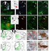
Figure 1
Figure 1. Glycinergic afferents in the mouse and human IL.
a) Injection site of the retrograde tracer fluorogold (FG) into the IL of a GlyT2::eGFP mouse. b-d) Retrogradely labeled GlyT2::eGFP-positive (arrows) and negative (arrowheads) neurons in the nucleus pontis oralis (PnO) at the coronal level indicated in the inset. e) Injection site of the anterograde tracer PHAL into the PnO and (f-g) anterogradely labeled GlyT2::eGFP-positive fibers (arrows) in the IL at the position shown in the inset. Distribution of GlyT2 fibers in the mouse (i,j) and human (m,n) thalamus at two coronal levels. The figures represent cumulative data. Light microscopic images of GlyT2-positive fibers and innervation of calbindin-positive cells via multiple contacts in the mouse (k, l) and human (o, p) IL. Scale bars: A,E, 1mm; all other 20 µm. CB, calbindin, For other abbreviations see Supplementary Fig 1,2.
Figure 2
Figure 2. Glycinergic terminals in IL are multisynaptic, co-express GABA and display variable postsynaptic receptor composition.
a) 3D reconstruction of a GlyT2::eGFP-positive terminal in IL from serial electron microscopic (EM) images, three of which are shown on the right. Green, synapses; magenta, puncta adherentia; dark blue, membrane of the terminal; light blue, glia; arrows, synapse; arrowheads puncta adherantia. b) Consecutive electron micrographs of a GlyT2:eGFP bouton (b) in the mouse IL immunostained for eGFP using preembedding silver staining (left), and for GABA using postembedding immunogold labeling (right). c) Comparison of random dendritic diameters (white bars) in the IL and the diameter of targets postsynaptic to GlyT2::eGFP terminals (black bars). Random dendrite diameters are also shown and as the ratio of summated perimeter of the dendrites in each bin (black line with diamonds), which better reflect the available membrane surfaces. d) Correlation between the synapse numbers of the GlyT2::eGFP boutons and the diameter of the postsynaptic IL dendrites. e) The average number of synapses with increasing distances from a given synapse in eGFP boutons in the IL. f) Electron micrograph of a GlyT2-immunopositive axon terminal in the human IL. green arrowheads, synapses g) White arrows point to colocalization of the γ2 subunit of GABAA receptors and of glycine receptors postsynaptic to GlyT2::eGFP terminals. The cityscape plot (h) represents the number of apposed GABAγ2R receptor and GlyR clusters per GlyT2::eGFP varicosity. Scales: a, b, f, 500 nm; g 1μm.
Figure 3
Figure 3. Glycinergic input evokes non-depressing inhibition and reduces IL cell firing.
a) Scheme of the experiment. b) ChR2-eYFP containing fibers in the IL. c) Averaged sample trace of light-evoked IPSCs before (black trace) and after (red) application of gabazine and strychnine. d) Variable mixed GABA/glycinergic phenotype of light-evoked IPSCs. Three different examples are shown with only GABAergic (top), mixed GABA/glycinergic (center), and only glycinergic (bottom) transmission. e) Ratio of IL cells showing various proportions of glycinergic leIPSCs. f-g ) SR95531 application leads to a significant acceleration of the decay time course of the leIPSCs. See the averaged traces for a single recorded cell in (f), and the pooled results for all the experiments in (g). h) Light-evoked responses display little depression during stimulation trains at different frequencies. i) Activation of GlyT2 fibers interrupts firing of IL neurons recorded in the current clamp configuration.
Figure 4
Figure 4. Activation of glycinergic afferents interrupts ongoing behavior activity.
a) Experimental design. b) Mice trajectory during the 1st (red) and 2nd (green) 5 s of optogenetic activation of GlyT2 fibers in the IL and during laser light shut off (black lines), in control (eYFP, left) and experimental (ChR2-eYFP, right) conditions. c) Average movement of control (dashed trace) and optogenetically activated (continuous trace) mice before, during (blue bar) and after stimulation. Error bars represent the s.e.m.
Figure 5
Figure 5. Activation of glycinergic afferents interrupts ongoing cortical activity.
a) Representative standardized frontal cortical LFP traces before (1) and during (2) the optogenetic activation of GlyT2 fibers in the IL. b) Wavelet spectrum of the cortical LFP showing the 33 s long activation period together with pre- and post-illumination period. Grey bars indicate the position of LFP samples in (a). Warm colors indicate higher power. c) Power spectra of the cortical LFPs in the 30 s preceding the stimulation (orange) and during photoactivation (blue) of the GlyT2 fibers in IL. d) Statistical comparison of the power spectra of the stimulated and control periods in one representative animal (n=25 stimulations). Gray bar indicates the frequency range which displayed statistically significant difference (2.14 Hz - 5.8 Hz, Mann-Whitney U test). In this range the highest p value was 0.00226 at 5.8 Hz (W=457). All other p values were lower. Error bars represent the s.e.m. au arbitrary unit.
Figure 6
Figure 6. Activity of GlyT2-positive neurons in the PRF in vivo is linked to cortical slow oscillation.
a) Experimental design. b) Spiking activity of a GlyT2 cell in vivo under ketamine-xylazine anesthesia (bottom trace) together with the cortical LFP (top trace) and filtered cortical multiunit activity (MUA, middle trace). c)The recorded and neurobiotin filled cell display GlyT2::eGFP expression. d) Neurolucida reconstruction of the recorded cell. e-f) Phase distribution of the firing activity of five different GlyT2::eGFP-positive neurons relative to the cortical slow oscillation. One cycle is 360o, 0o peak of the UP state. Note the different phase preference of each cell. Scale: c, 20 µm; d, 100 µm.

References

    1. Drew T, Andujar J-E, Lajoie K, Yakovenko S. Cortical mechanisms involved in visuomotor coordination during precision walking. Brain Res Rev. 2008;57:199–211. - PubMed
    1. Lemon RN. Descending pathways in motor control. Annu Rev Neurosci. 2008;31:195–218. - PubMed
    1. Rizzolatti G, Luppino G. The cortical motor system. Neuron. 2001;31:889–901. - PubMed
    1. Graybiel AM. Habits, rituals, and the evaluative brain. Annu Rev Neurosci. 2008;31:359–87. - PubMed
    1. Isoda M, Hikosaka O. Cortico-basal ganglia mechanisms for overcoming innate, habitual and motivational behaviors. Eur J Neurosci. 2011;33:2058–69. - PMC - PubMed

Publication types

LinkOut - more resources