

Enhanced invasion of metastatic cancer cells via extracellular matrix interface
- PMID: 25706718
- PMCID: PMC4338181
- DOI: 10.1371/journal.pone.0118058
Enhanced invasion of metastatic cancer cells via extracellular matrix interface
Abstract
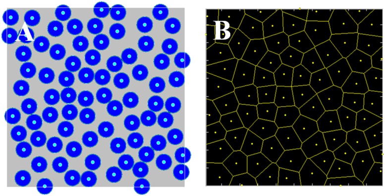
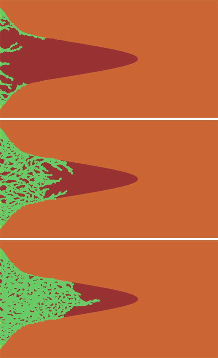
Cancer cell invasion is a major component of metastasis and is responsible for extensive cell diffusion into and major destruction of tissues. Cells exhibit complex invasion modes, including a variety of collective behaviors. This phenomenon results in the structural heterogeneity of the extracellular matrix (ECM) in tissues. Here, we systematically investigated the environmental heterogeneity facilitating tumor cell invasion via a combination of in vitro cell migration experiments and computer simulations. Specifically, we constructed an ECM microenvironment in a microfabricated biochip and successfully created a three-dimensional (3D) funnel-like matrigel interface inside. Scanning electron microscopy demonstrated that the interface was at the interior defects of the nano-scale molecular anisotropic orientation and the localized structural density variations in the matrigel. Our results, particularly the correlation of the collective migration pattern with the geometric features of the funnel-like interface, indicate that this heterogeneous in vitro ECM structure strongly guides and promotes aggressive cell invasion in the rigid matrigel space. A cellular automaton model was proposed based on our experimental observations, and the associated quantitative analysis indicated that cell invasion was initiated and controlled by several mechanisms, including microenvironment heterogeneity, long-range cell-cell homotype and gradient-driven directional cellular migration. Our work shows the feasibility of constructing a complex and heterogeneous in vitro 3D ECM microenvironment that mimics the in vivo environment. Moreover, our results indicate that ECM heterogeneity is essential in controlling collective cell invasive behaviors and therefore determining metastasis efficiency.
Conflict of interest statement
Figures


Similar articles
-
Modeling the cholesteatoma microenvironment: coculture of HaCaT keratinocytes with WS1 fibroblasts induces MMP-2 activation, invasive phenotype, and proteolysis of the extracellular matrix.Laryngoscope. 2007 Feb;117(2):313-8. doi: 10.1097/01.mlg.0000251164.26405.1a. Laryngoscope. 2007. PMID: 17204986
-
PTEN deletion potentiates invasion of colorectal cancer spheroidal cells through 3D Matrigel.Integr Biol (Camb). 2015 Mar;7(3):324-34. doi: 10.1039/c4ib00298a. Epub 2015 Jan 27. Integr Biol (Camb). 2015. PMID: 25625883
-
Influence of a self-assembling peptide, RADA16, compared with collagen I and Matrigel on the malignant phenotype of human breast-cancer cells in 3D cultures and in vivo.Macromol Biosci. 2009 May 13;9(5):437-43. doi: 10.1002/mabi.200800262. Macromol Biosci. 2009. PMID: 19165822
-
The matrix environmental and cell mechanical properties regulate cell migration and contribute to the invasive phenotype of cancer cells.Rep Prog Phys. 2019 Jun;82(6):064602. doi: 10.1088/1361-6633/ab1628. Epub 2019 Apr 4. Rep Prog Phys. 2019. PMID: 30947151 Review.
-
Cell-ECM Interactions in Tumor Invasion.Adv Exp Med Biol. 2016;936:73-91. doi: 10.1007/978-3-319-42023-3_4. Adv Exp Med Biol. 2016. PMID: 27739043 Review.
Cited by
-
Circulating Tumor Cells as a Tool for Assessing Tumor Heterogeneity.Theranostics. 2019 Jun 19;9(16):4580-4594. doi: 10.7150/thno.34337. eCollection 2019. Theranostics. 2019. PMID: 31367241 Free PMC article. Review.
-
Towards organoid culture without Matrigel.Commun Biol. 2021 Dec 10;4(1):1387. doi: 10.1038/s42003-021-02910-8. Commun Biol. 2021. PMID: 34893703 Free PMC article. Review.
-
T lymphocytes against solid malignancies: winning ways to defeat tumours.Cell Stress. 2018 Jul 26;2(8):200-212. doi: 10.15698/cst2018.07.148. Cell Stress. 2018. PMID: 31225487 Free PMC article. Review.
-
Three-Dimensional Culture System of Cancer Cells Combined with Biomaterials for Drug Screening.Cancers (Basel). 2020 Sep 24;12(10):2754. doi: 10.3390/cancers12102754. Cancers (Basel). 2020. PMID: 32987868 Free PMC article. Review.
-
The emerging role of circulating tumor cells in cancer management.Am J Transl Res. 2020 Feb 15;12(2):332-342. eCollection 2020. Am J Transl Res. 2020. PMID: 32194887 Free PMC article. Review.
References
-
- Sahai E. Mechanisms of cancer cell invasion. Current Opinion in Genetics & Development. 2005;15: 87–96. - PubMed
Publication types
MeSH terms
Substances
LinkOut - more resources
Full Text Sources
Other Literature Sources
