. 2015 Feb 23;10(2):e0118684.
doi: 10.1371/journal.pone.0118684. eCollection 2015.

How structural and physicochemical determinants shape sequence constraints in a functional enzyme

Affiliations

How structural and physicochemical determinants shape sequence constraints in a functional enzyme

Luciano A Abriata et al. PLoS One. .

Abstract

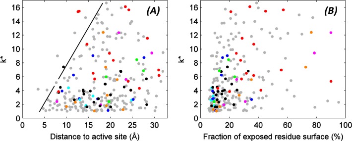
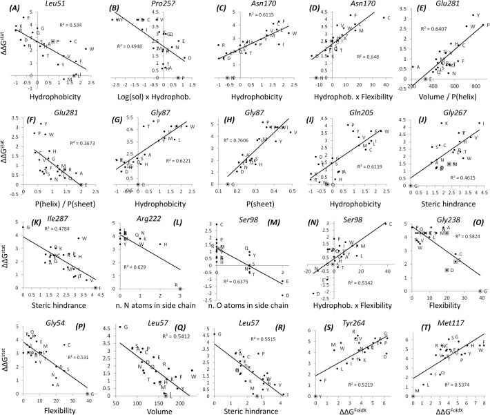
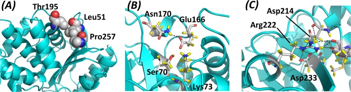
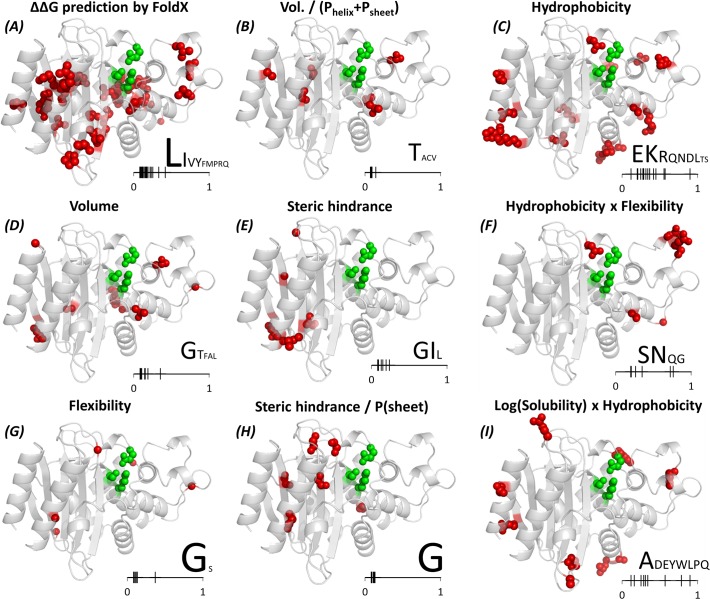
The need for interfacing structural biology and biophysics to molecular evolution is being increasingly recognized. One part of the big problem is to understand how physics and chemistry shape the sequence space available to functional proteins, while satisfying the needs of biology. Here we present a quantitative, structure-based analysis of a high-resolution map describing the tolerance to all substitutions in all positions of a functional enzyme, namely a TEM lactamase previously studied through deep sequencing of mutants growing in competition experiments with selection against ampicillin. Substitutions are rarely observed within 7 Å of the active site, a stringency that is relaxed slowly and extends up to 15-20 Å, with buried residues being especially sensitive. Substitution patterns in over one third of the residues can be quantitatively modeled by monotonic dependencies on amino acid descriptors and predictions of changes in folding stability. Amino acid volume and steric hindrance shape constraints on the protein core; hydrophobicity and solubility shape constraints on hydrophobic clusters underneath the surface, and on salt bridges and polar networks at the protein surface together with charge and hydrogen bonding capacity. Amino acid solubility, flexibility and conformational descriptors also provide additional constraints at many locations. These findings provide fundamental insights into the chemistry underlying protein evolution and design, by quantitating links between sequence and different protein traits, illuminating subtle and unexpected sequence-trait relationships and pinpointing what traits are sacrificed upon gain-of-function mutation.


Conflict of interest statement

Competing Interests: The authors have declared that no competing interests exist.

Figures

Fig 1
Fig 1. Effective number of substitutions versus distance to the active site and solvent exposure.
Effective number of amino acids that appear at each position (k*) plotted versus the distance from its Cα atom to the active site (A) and against the fraction of amino acid surface exposed to the solvent (B). The position of the active site was defined as the average position of the Ser70, Lys73 and Glu166 Cα atoms; surface exposure was computed with the POPS webserver [48,49] based on the X-ray structure deposited under PDB entry 1XPB [50]. Dot colors correspond to correlations against the most often matched properties: blue for volume, cyan for volume/(P(helix)+P(sheet)), green for steric hindrance, dark green for steric hindrance / P(sheet), red for hydrophobicity, magenta for log(solubility) x hydrophobicity, orange for flexibility x hydrophobicity. Black dots represent correlations with FoldX predictions. Gray dots correspond to the rest of the residues (with other or no detected correlations). The line drawn in panel A arbitrarily indicates how the maximum possible k* increases with distance to the active site.
Fig 2
Fig 2. Examples of correlations detected between ΔΔGstat values and amino acid descriptors (a-r) or ΔΔGFoldX (s-t).
The gray circles point at the wild type amino acid, gray squares point at substitutions that have been observed in natural TEM variants. Lines correspond to best linear fits.
Fig 3
Fig 3. Structural representation of the (a) Leu51 (b) Asn170 (c) Arg222 regions in the TEM-1 β-lactamase structure.
Pictures rendered from PDB ID 1XPB [50] using the program PyMOL [51]. Atom colors are red for oxygen, blue for nitrogen, gray for carbon and yellow for hydrogen.
Fig 4
Fig 4. Structure mapping of the residues whose substitution patterns can be explained by the nine most common descriptors.
The mapped amino acids are shown as red spheres, and residues Ser70, Lys73 and Glu166 as green spheres. All residue representations lack the main chain nitrogen, carbonyl carbon and oxygen atoms for clarity. The letters on the bottom right of each panel indicate the wild type amino acids most often found at the indicated locations, with the font size being roughly proportional to the relative number of occurrences of the amino acid. The small bar on the bottom right of each panel measures the fractional solvent exposure of the wild type residues to which the descriptor was mapped.

References

    1. Aharoni A, Gaidukov L, Khersonsky O, McQ Gould S, Roodveldt C, et al. (2005) The “evolvability” of promiscuous protein functions. Nat Genet 37, 73–76 - PubMed
    1. Thorne JL (2007) Protein evolution constraints and model-based techniques to study them. Curr Opin Struct Biol 17, 337–341 - PubMed
    1. Tokuriki N, Tawfik DS (2009) Stability effect of mutations and protein evolvability. Curr Opin Struct Biol 19, 596–604 10.1016/j.sbi.2009.08.003 - DOI - PubMed
    1. Fornasari MS, Parisi G, Echave J (2007) Quaternary structure constraints on evolutionary sequence divergence. Mol Biol Evol 24, 349–351 - PubMed
    1. Meyer AG, Wilke CO (2013) Integrating sequence variation and protein structure to identify sites under selection. Mol Biol Evol 30, 36–44 10.1093/molbev/mss217 - DOI - PMC - PubMed

Publication types

Substances

LinkOut - more resources