Review
. 2015:84:519-50.
doi: 10.1146/annurev-biochem-060614-034411. Epub 2015 Feb 20.

Natural photoreceptors as a source of fluorescent proteins, biosensors, and optogenetic tools

Affiliations
Review

Natural photoreceptors as a source of fluorescent proteins, biosensors, and optogenetic tools

Daria M Shcherbakova et al. Annu Rev Biochem. 2015.

Abstract

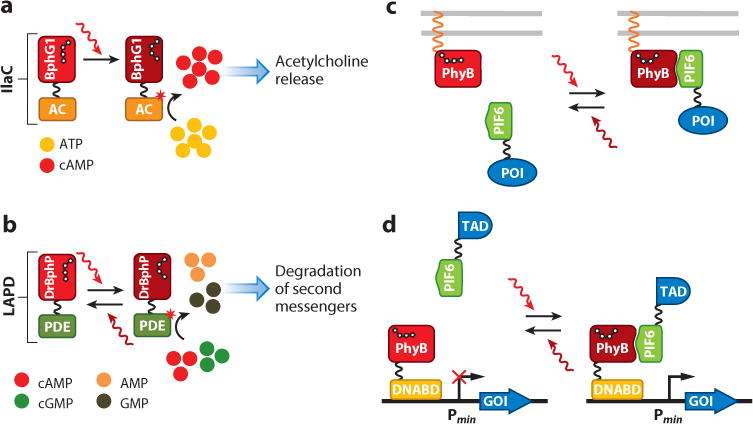
Genetically encoded optical tools have revolutionized modern biology by allowing detection and control of biological processes with exceptional spatiotemporal precision and sensitivity. Natural photoreceptors provide researchers with a vast source of molecular templates for engineering of fluorescent proteins, biosensors, and optogenetic tools. Here, we give a brief overview of natural photoreceptors and their mechanisms of action. We then discuss fluorescent proteins and biosensors developed from light-oxygen-voltage-sensing (LOV) domains and phytochromes, as well as their properties and applications. These fluorescent tools possess unique characteristics not achievable with green fluorescent protein-like probes, including near-infrared fluorescence, independence of oxygen, small size, and photosensitizer activity. We next provide an overview of available optogenetic tools of various origins, such as LOV and BLUF (blue-light-utilizing flavin adenine dinucleotide) domains, cryptochromes, and phytochromes, enabling control of versatile cellular processes. We analyze the principles of their function and practical requirements for use. We focus mainly on optical tools with demonstrated use beyond bacteria, with a specific emphasis on their applications in mammalian cells.

Keywords: BphP; CRY2; LOV domain; bacteriophytochrome; iRFP; optogenetics; phytochrome.


Figures

Figure 1
Figure 1
A variety of photoreceptors and their chromophores explored as templates to engineer optical tools. (Left) Chemical structures of the chromophores and a color-scale bar representing the wavelength range of their absorbance in natural photoreceptors. The chromophores are presented in their inactive (dark) states. The retinal structure corresponds to microbial opsins. For each chromophore, its primary photochemistry is indicated. An asterisk indicates the C4a atom in FMN, which forms a covalent bond with a conservative cysteine in LOV domains upon light illumination. The arrow in some chemical structures indicates the double bond that isomerizes upon activation. (Right) Natural photoreceptors that have been actively explored to develop genetically encoded optical tools. For each type of photoreceptor, a schematic domain structure of one example receptor is presented. Note that all photoreceptors, except opsins, have a modular domain organization with separate photosensory module and effector domain. Abbreviations: AMP, adenosine 5′-monophosphate; Asphot1, Avena sativa phototropin 1; AtCRY2, Arabidopsis thaliana cryptochrome 2; AtPhyB, A. thaliana phytochrome B; BLUF, blue-light-utilizing flavin adenine dinucleotide; CrChR2, Chlamydomonas reinhardtii channelrhodopsin-2; DrBphP, Deinococcus radiodurans bacterial phytochrome; LOV, light-oxygen-voltage-sensing.
Figure 2
Figure 2
Engineering of FPs from photoreceptors and their advantages over GFP-like FPs. (a) Strategy for engineering of LOV domain–based FPs. The truncated LOV domain is subjected to molecular evolution to block FMN–cysteinyl adduct formation and improve brightness and chromophore incorporation. (b) LOV domain–based FPs are small (~10–15 kDa) and do not require molecular oxygen for maturation. Thus, they can be utilized in applications as small fusion tags and as reporters in anaerobic conditions. In the presence of oxygen, LOV domain–based FPs generate reactive oxygen species (singlet oxygen) and, thus, can serve as photosensitizers for photodestruction of proteins and cells. (c) Strategy for engineering of permanently fluorescent and photoswitchable NIR FPs. To abolish photoswitching and obtain a permanently fluorescent FP, the phytochrome is truncated to chromophore-binding PAS and GAF domains. Directed molecular evolution allows one to improve the brightness, efficiency, and specificity of BV incorporation. For development of photoactivatable FPs, the PHY domain is not deleted to preserve photoswitchable properties. In molecular evolution, variants that do not photoswitch from fluorescent Pr form under weak illumination during imaging are selected. (d) The main advantage of FPs derived from bacterial phytochromes is their NIR-shifted spectra. In the NIR transparency window (650–900 nm), mammalian tissues are most transparent to light because combined absorption of hemoglobin and water is minimal. Thus, NIR FPs are suitable for noninvasive whole-body imaging. Abbreviations: FMN, flavin mononucleotide; FP, fluorescent protein; GAF, cGMP phosphodiesterase/adenylate cyclase/FhlA; GFP, green fluorescent protein; LOV, light-oxygen-voltage-sensing; NIR, near-infrared; PAS, Per–Arnt–Sim; PHY, phytochrome-specific; POI, protein of interest.
Figure 3
Figure 3
Fluorescent reporters designed from photoreceptors. (a) A LOV-based reporter for oxygen. In anaerobic conditions, YFP does not form a chromophore, and no FRET between LOV-based FP and YFP occurs. In the presence of oxygen, a YFP chromophore is formed, and the matured YFP acts as a FRET acceptor for the LOV-based FP. The process is not reversible. Thus, intracellular stability and turnover of reporter influence its response and its kinetics. (b) Phytochrome-based reporters for protein–protein interactions using a bimolecular fluorescence complementation approach. Upon interaction of two protein partners, the fused PAS and GAF phytochrome domains complement into a functional FP. Abbreviations: FP, fluorescent protein; FRET, Förster resonance energy transfer; GAF, cGMP phosphodiesterase/adenylate cyclase/FhlA; LOV, light-oxygen-voltage-sensing; PAS, Per–Arnt–Sim; YFP, yellow fluorescent protein.
Figure 4
Figure 4
General strategies to engineer optogenetic tools. (a) Caging the protein interaction interface/enzyme active site inhibits protein activity in darkness. During light absorption, photoreceptors or light-sensitive domains can undergo structural rearrangements and uncage the interface/active site of the caged protein in the lit state. (b) Light-induced structural changes in the photosensory core may lead to tertiary structure perturbations in the whole protein molecule and activate effector domain. (c,d) After light illumination, the quaternary structure of photoreceptors can change. They can (c) hetero-oligomerize or (d) homo-oligomerize and may control interaction of fused protein partners, reconstitute split enzymes, or relocalize fused proteins to other cell compartments. The term hv designates the activation light. The red asterisk indicates an activated state.
Figure 5
Figure 5
Optogenetic constructs that are activated by blue light. (a) After illumination of asLOV2–Rac1 [Avena sativa LOV (light-oxygen-voltage-sensing) domain 2] fusion, FMN (flavin mononucleotide) binding occurs, and Rac1, which was caged on the asLOV2 core, is released. This process induces lamellipodia formation through actin polymerization. (b) TULIP (tunable light-induced dimerization tags) technology is based on interaction of the ePDZ (enhanced postsynaptic density protein 95, disc-large tumor suppressor protein, zonula occludens 1) domain with its binding epitope caged on the asLOV2 core in the dark state. Illumination induces structural changes in asLOV2, and unwinding of the Jα helix results in the availability of a binding epitope for ePDZ. Thus, a protein fused to the ePDZ domain can be translocated to a specific cell compartment and perform its function. (c) Light-induced interaction between GIGANTEA (GI) and FKF1 (flavin-binding kelch repeat F-box 1) proteins and their heterodimerization result in lamellipodia formation through attraction of Rac1 fused with FKF1 to the plasma membrane. (d) For light-induced transcription activation, GI is fused to the DNA-binding domain (Gal4) and FKF1 is fused to the transcription activation domain (TAD). In the lit state, GI interacts with FKF1, and transcription activation by TAD occurs from the minimal promoter (Pmin). (e) A protein of interest (POI) can be recruited to the plasma membrane by fusing it to cryptochrome 2 (CRY2), which interacts with its membrane-anchored partner CIB1 [CRY-interacting bHLH1 (helix-loop-helix 1)] upon illumination. (f) After illumination, reconstitution of split Cre recombinase occurs when the N-terminal part of Cre is fused to CRY2 and the C-terminal part of Cre is fused to CIB1. Reconstituted Cre removes the terminator region (STOP) flanked by loxP sites and activates reporter gene transcription from the constitutive promoter (Pconst). (g) Cell morphology can be controlled by light-induced clustering of Vav2, a guanine nucleotide exchange factor that activates Rho small GTPases. In the dark state, the Vav2–CRY2 fusion is localized near the plasma membrane and induces protrusion formation. In the lit state, CRY2 interacts with CIB1 fused with the multimeric protein (MP), and formation of large clusters occurs, thus inhibiting Vav2 in clusters and causing retraction of the plasma membrane. The term hv designates the activation light.
Figure 6
Figure 6
Optogenetic tools based on phytochromes. (a) Near-infrared (NIR) light–activated adenylate cyclase (IlaC) consists of a photosensory module of Rhodobacter sphaeroides BphP (bacterial phytochrome), BphG1, and the AC (adenylate cyclase) domain from Nostoc sp. CyaB1. Synthesis of cAMP by IlaC can be activated with NIR light. When expressed in cholinergic neurons of Caenorhabditis elegans, it affects worm behavior in a light-dependent manner by increasing intracellular cAMP, which leads to release of acetylcholine and subsequent activation of muscles. (b) Light-activated phosphodiesterase (LAPD) is designed by fusing the photosensory module of Deinococcus radiodurans BphP with the effector domain of human phosphodiesterase 2A (PDE). Upon NIR illumination, phosphodiesterase becomes active and upregulates hydrolysis of second messengers, such as cAMP and cGMP. (c) Light-controllable reversible interaction of PhyB and phytochrome-interacting factor 6 (PIF6) can be exploited for recruitment of the protein of interest (POI) to the plasma membrane. After 650-nm light illumination, PhyB undergoes structural rearrangements and interacts with PIF6. This interaction is reversible, and dissociation can be activated by 750-nm light. (d) Light-induced heterodimerization can be used for transcription activation of the gene of interest (GOI) from the minimal promoter (Pmin). PhyB is fused to the DNA-binding domain (DNABD), and PIF6 is fused to the transcription activation domain (TAD). Upon 660-nm light illumination, PhyB and PIF6 interact, and TAD activates transcription from the Pmin. Dissociation of the PhyB–PIF6 pair can be activated by 740-nm light, and the system can be reversibly toggled between the stable on (transcription is activated) and off (transcription is terminated) states. The asterisk indicates an activated state.

References

    1. van der Horst MA, Hellingwerf KJ. Photoreceptor proteins, “star actors of modern times”: a review of the functional dynamics in the structure of representative members of six different photoreceptor families. Acc Chem Res. 2004;37:13–20. - PubMed
    1. Christie JM, Gawthorne J, Young G, Fraser NJ, Roe AJ. LOV to BLUF: flavoprotein contributions to the optogenetic toolkit. Mol Plant. 2012;5:533–44. - PubMed
    1. Rockwell NC, Lagarias JC. A brief history of phytochromes. ChemPhysChem. 2010;11:1172–80. - PMC - PubMed
    1. Losi A, Gärtner W. The evolution of flavin-binding photoreceptors: an ancient chromophore serving trendy blue-light sensors. Annu Rev Plant Biol. 2012;63:49–72. - PubMed
    1. Chaves I, Pokorny R, Byrdin M, Hoang N, Ritz T, et al. The cryptochromes: blue light photoreceptors in plants and animals. Annu Rev Plant Biol. 2011;62:335–64. - PubMed

Publication types

LinkOut - more resources