. 2015 Feb 23;10(2):e0118286.
doi: 10.1371/journal.pone.0118286. eCollection 2015.

Phenotypic responses of differentiated asthmatic human airway epithelial cultures to rhinovirus

Affiliations

Phenotypic responses of differentiated asthmatic human airway epithelial cultures to rhinovirus

Jianwu Bai et al. PLoS One. .

Abstract

Objectives: Human airway epithelial cells are the principal target of human rhinovirus (HRV), a common cold pathogen that triggers the majority of asthma exacerbations. The objectives of this study were 1) to evaluate an in vitro air liquid interface cultured human airway epithelial cell model for HRV infection, and 2) to identify gene expression patterns associated with asthma intrinsically and/or after HRV infection using this model.

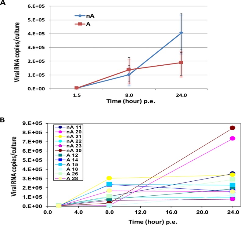
Methods: Air-liquid interface (ALI) human airway epithelial cell cultures were prepared from 6 asthmatic and 6 non-asthmatic donors. The effects of rhinovirus RV-A16 on ALI cultures were compared. Genome-wide gene expression changes in ALI cultures following HRV infection at 24 hours post exposure were further analyzed using RNA-seq technology. Cellular gene expression and cytokine/chemokine secretion were further evaluated by qPCR and a Luminex-based protein assay, respectively.

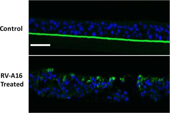
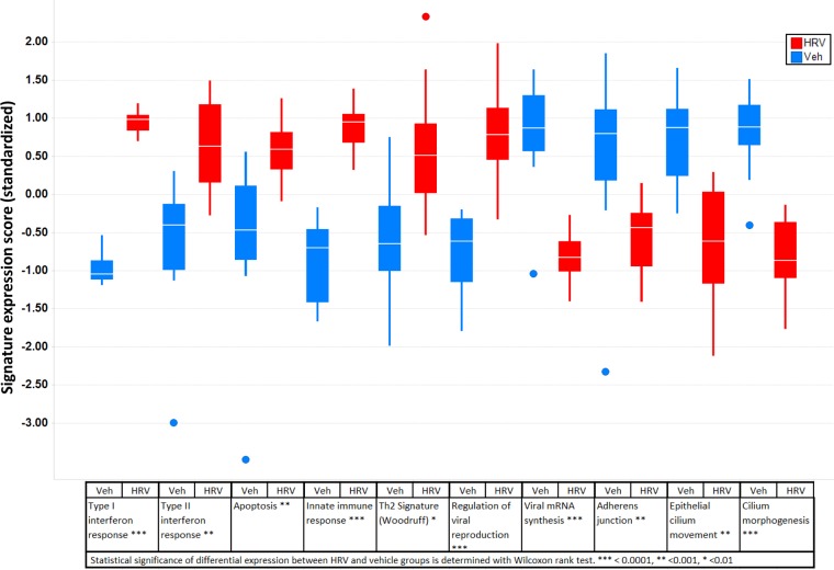
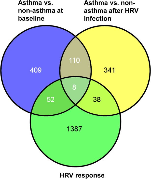
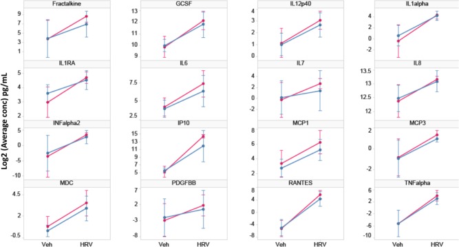
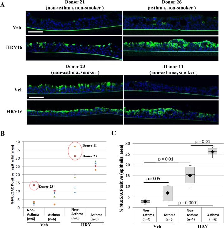
Main results: ALI cultures were readily infected by HRV. RNA-seq analysis of HRV infected ALI cultures identified sets of genes associated with asthma specific viral responses. These genes are related to inflammatory pathways, epithelial structure and remodeling and cilium assembly and function, including those described previously (e.g. CCL5, CXCL10 and CX3CL1, MUC5AC, CDHR3), and novel ones that were identified for the first time in this study (e.g. CCRL1).

Conclusions: ALI-cultured human airway epithelial cells challenged with HRV are a useful translational model for the study of HRV-induced responses in airway epithelial cells, given that gene expression profile using this model largely recapitulates some important patterns of gene responses in patients during clinical HRV infection. Furthermore, our data emphasize that both abnormal airway epithelial structure and inflammatory signaling are two important asthma signatures, which can be further exacerbated by HRV infection.


Conflict of interest statement

Competing Interests: This work was supported by Merck Research Laboratories and MatTek. Co-authors Jianwu Bai, Steven L. Smock, Kenzie D. MacIsaac, Yongsheng Huang, Brian Roberts, Yu-Lu Ma, Joel A. Klappenbach, Michael A. Crackower and Stephen E. Alves are employed by Merck Research Laboratories. Co-authors George R. Jackson, Jr, Courtney Mankus, Jonathan Oldach and Patrick J. Hayden are employed by MatTek Corporation. There are no patents, products in development or marketed products to declare. Although MatTek produces commercial ALI culture products, all methods are disclosed to allow independent production of the models described in the paper. This does not alter the authors’ adherence to all the PLOS ONE policies on sharing data and materials.

Figures

Fig 1
Fig 1. Time course of RV-A16 infection for asthmatic (n = 6) and non-asthmatic (n = 6) donor groups (A) and each donor (B) in ALI cultures.
Viral genome levels in cell lysates were measured by qPCR at three time points, and were presented as viral RNA copies /culture. There was no significant difference in the mean viral genome levels between asthmatic and non-asthmatic samples. The p values between asthma and non-asthmatic samples are 0.48, 0.53 and 0.13 at 1.5, 8, and 24 hours post exposure (p.e.) respectively.
Fig 2
Fig 2. ALI cultured HAECs were infectible by RV-A16.
Immunofluorescence micrographs of transverse sections of ALI cultures shows RV-A16 replicated in ALI cultured HAECs. Positive immunofluorescence staining for mabj2 (in green) was not observed in uninfected control tissues (top panel), but was observed in RV-A16 infected tissues (bottom panel); DAPI staining in blue shows cell nuclei in the tissues. Scale bars: 50 μm.
Fig 3
Fig 3. Functional pathways that showed statistically significant differential expression between RV-A16 and Vehicle groups (p<0.05).
Outliner is shown individually with a solid circle.
Fig 4
Fig 4. Distributions of differentially expressed genes in asthmatic samples after treatment of vehicle or RV-A16.
579 genes in the “Asthma vs. non-asthma at baseline” group had baseline difference between asthmatic and non-asthmatic cells (p<0.05); 497 genes in the “Asthma vs. non-asthma after HRV infection” group had asthma-related viral response (p<0.05); and 1485 genes in the “HRV response group had strong expression changes associated with viral infection across both asthma and non-asthma donors. The numbers of genes only in one group or common for either two or three groups are shown by either exclusive or overlapping areas in Venn diagrams. The diagrams were generated with an online tool available at http://bioinfogp.cnb.csic.es/tools/venny/.
Fig 5
Fig 5. Expression of secreted cytokines/chemokines in culture medium.
16/42 cytokines tested had robust expression levels and were significantly up-regulated at 24 hr after RV-A16 infection across 12 donors (p value<0.05). Differences were not significant between asthma and non-asthma groups (p>0.05). Red lines represent asthma samples, blue lines represent non-asthma samples. Each line represents the mean and standard deviation of log2 concentration of each cytokine/chemokine.
Fig 6
Fig 6. MUC5AC expression is upregulated in response to HRV infection.
A) Immunofluorescence of MUC5AC in ALI cultures from donors 21 (non-asthma, non-smoker), 23 (non-asthma, smoker), 26 (asthma, non-smoker) and 11(asthma, smoker) show an increase in staining with HRV treatment, with only donor 23 having high baseline expression. Green: MUC5AC, blue: DAPI staining for nuclei, representative images, scale bar: 50 μm. N = 6 ALI cultures per group (asthma and non-asthma). B and C) Quantification of immunofluorescence staining presented as either dot plot (B) or box plot (C). Positively stained area was measured and presented as % of total epithelial area. Red dots indicate outliers (> 1.5x inter-quartile distance). B) Inclusion of all data from 6 non-asthmatic and 6 asthmatic donors in analysis. C) Removal of non-diseased smokers (donors 11 and 23) from the data set results in significant differences between HRV and vehicle treated tissues in both non-asthmatic and asthmatic donors as determined by ANOVA and Tukey HSD test.

References

    1. Hershenson MB. Rhinovirus-Induced Exacerbations of Asthma and COPD. Scientifica (Cairo) 2013: 405876 10.1155/2013/405876 - DOI - PMC - PubMed
    1. Gavala ML, Bertics PJ, Gern JE. Rhinoviruses, allergic inflammation, and asthma. Immunol Rev. 2011; 242: 69–90. 10.1111/j.1600-065X.2011.01031.x - DOI - PMC - PubMed
    1. Gern JE. The ABCs of rhinoviruses, wheezing, and asthma. J Virol. 2010; 84: 7418–7426. 10.1128/JVI.02290-09 - DOI - PMC - PubMed
    1. Holgate ST. The sentinel role of the airway epithelium in asthma pathogenesis. Immunol Rev. 2011; 242: 205–219. 10.1111/j.1600-065X.2011.01030.x - DOI - PubMed
    1. Holgate ST. Mechanisms of Asthma and Implications for Its Prevention and Treatment: A Personal Journey. Allergy Asthma Immunol Res. 2013; 5: 343–347. 10.4168/aair.2013.5.6.343 - DOI - PMC - PubMed

Publication types

MeSH terms