

Bilateral central retinal artery occlusion associated with herpes simplex virus-associated acute retinal necrosis and meningitis: case report and literature review
- PMID: 25707059
- PMCID: PMC4557211
- DOI: 10.3928/23258160-20150213-24
Bilateral central retinal artery occlusion associated with herpes simplex virus-associated acute retinal necrosis and meningitis: case report and literature review
Abstract
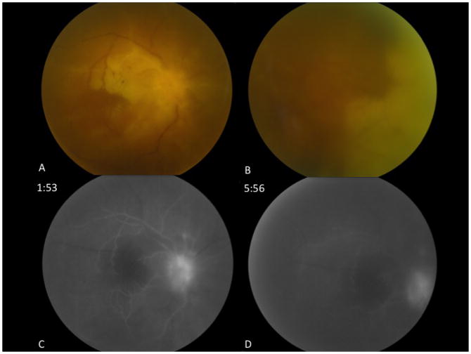
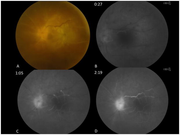
A 60-year-old woman with a history of recurrent headaches and blurred vision presented with bilateral optic disc edema. Optic neuritis was suspected, and intravenous methylprednisonlone was administered. Her vision declined to hand motions in both eyes, and subsequent evaluation revealed bilateral acute retinal necrosis with bilateral central retinal artery occlusions (CRAO). Aqueous humor polymerase chain reaction analysis was positive for herpes simplex virus (HSV), establishing a diagnosis of HSV-associated bilateral acute retinal necrosis (ARN) and meningitis. CRAO has rarely been reported in association with ARN, and a fulminant course with bilateral CRAO in association with ARN has not been previously reported. This case emphasizes the importance of careful peripheral examination in patients with presumptive optic neuritis, judicious use of systemic corticosteroid in this context, and the retinal vaso-obliterative findings that may be observed in the pathogenesis of ARN.
Copyright 2015, SLACK Incorporated.
Figures
Similar articles
-
Clinical characteristics of acute HSV-2 retinal necrosis.Am J Ophthalmol. 2004 May;137(5):872-9. doi: 10.1016/j.ajo.2003.12.036. Am J Ophthalmol. 2004. PMID: 15126152 Review.
-
Acute retinal necrosis secondary to herpes simplex virus type 2 in neonates.Ophthalmic Surg Lasers Imaging Retina. 2015 Apr;46(4):499-501. doi: 10.3928/23258160-20150422-18. Ophthalmic Surg Lasers Imaging Retina. 2015. PMID: 25932732
-
Herpes simplex virus-2 quantification by real-time polymerase chain reaction in acute retinal necrosis.Jpn J Ophthalmol. 2005 Jan-Feb;49(1):64-5. doi: 10.1007/s10384-004-0145-0. Jpn J Ophthalmol. 2005. PMID: 15692781 No abstract available.
-
Herpes simplex virus type 2 mediated acute retinal necrosis in a pediatric population: case series and review.Graefes Arch Clin Exp Ophthalmol. 2013 Feb;251(2):559-66. doi: 10.1007/s00417-012-2164-8. Epub 2012 Oct 5. Graefes Arch Clin Exp Ophthalmol. 2013. PMID: 23052715 Review.
-
Acute retinal necrosis secondary to herpes simplex virus type 2 with preexisting chorioretinal scarring.J Pediatr Ophthalmol Strabismus. 2008 Jan-Feb;45(1):59-61. doi: 10.3928/01913913-20080101-17. J Pediatr Ophthalmol Strabismus. 2008. PMID: 18286969
Cited by
-
Cytopathology of Vitreous Specimens in Acute Retinal Necrosis.Ocul Immunol Inflamm. 2022 Oct-Nov;30(7-8):1609-1616. doi: 10.1080/09273948.2021.1922926. Epub 2021 Jul 9. Ocul Immunol Inflamm. 2022. PMID: 34242097 Free PMC article.
-
Rare cause of left eye floaters and blurred vision in an immunocompromised patient: acute retinal necrosis (ARN).BMJ Case Rep. 2022 Mar 2;15(3):e248636. doi: 10.1136/bcr-2021-248636. BMJ Case Rep. 2022. PMID: 35236705 Free PMC article.
-
Advances in the Diagnosis and Management of Acute Retinal Necrosis.Ann Eye Sci. 2020 Sep;5:28. doi: 10.21037/aes-2019-dmu-09. Epub 2020 Sep 15. Ann Eye Sci. 2020. PMID: 33381683 Free PMC article.
-
Successful treatment with repeated dexamethasone implant injections for recurrent macular edema after acute retinal necrosis.J Ophthalmic Inflamm Infect. 2022 Oct 21;12(1):33. doi: 10.1186/s12348-022-00310-5. J Ophthalmic Inflamm Infect. 2022. PMID: 36269441 Free PMC article.
-
Outcomes of Combination Systemic and Intravitreal Antiviral Therapy for Acute Retinal Necrosis.Ophthalmol Retina. 2021 Mar;5(3):292-300. doi: 10.1016/j.oret.2020.07.012. Epub 2020 Jul 16. Ophthalmol Retina. 2021. PMID: 32683108 Free PMC article.
References
-
- Urayama A, Yamada N, Sasaki T. Unilateral acute uveitis with retinal periarteritis and detachment. Jpn J Clin Ophthalmol. 1971;(25):607–619.
-
- Ganatra JB, Chandler D, Santos C, Kuppermann B, Margolis TP. Viral causes of the acute retinal necrosis syndrome. Am J Ophthalmol. 2000;129(2):166–172. - PubMed
-
- Holland GN. Standard diagnostic criteria for the acute retinal necrosis syndrome. Executive Committee of the American Uveitis Society. Am J Ophthalmol. 1994;117(5):663–667. - PubMed
-
- Kang SW, Kim SK. Optic neuropathy and central retinal vascular obstruction as initial manifestations of acute retinal necrosis. Jpn J Ophthalmol. 2001;45(4):425–428. - PubMed
Publication types
MeSH terms
Substances
Grants and funding
LinkOut - more resources
Full Text Sources
Other Literature Sources
Medical
