

The Rheumatoid Arthritis Risk Gene LBH Regulates Growth in Fibroblast-like Synoviocytes
- PMID: 25707478
- PMCID: PMC4490933
- DOI: 10.1002/art.39060
The Rheumatoid Arthritis Risk Gene LBH Regulates Growth in Fibroblast-like Synoviocytes
Abstract
Objective: Fibroblast-like synoviocytes (FLS) are key players in the synovial pathology of rheumatoid arthritis (RA). Currently, there is no treatment that specifically targets these aggressive cells. By combining 3 different "omics" data sets, i.e., 1) risk genes in RA, 2) differentially expressed genes, and 3) differential DNA methylation in RA versus osteoarthritis (OA) FLS, we identified LBH (limb bud and heart development) as a candidate gene in RA. The present study was undertaken to define the role of this gene in FLS.
Methods: Synovial tissue specimens from RA and OA patients were collected at the time of joint replacement surgery. LBH expression was silenced using small interfering RNA or overexpressed using an LBH expression vector in primary FLS. Gene expression profiles were determined by microarray and assessed using Ingenuity Pathway Analysis. Effects of modified LBH expression were investigated in functional assays.
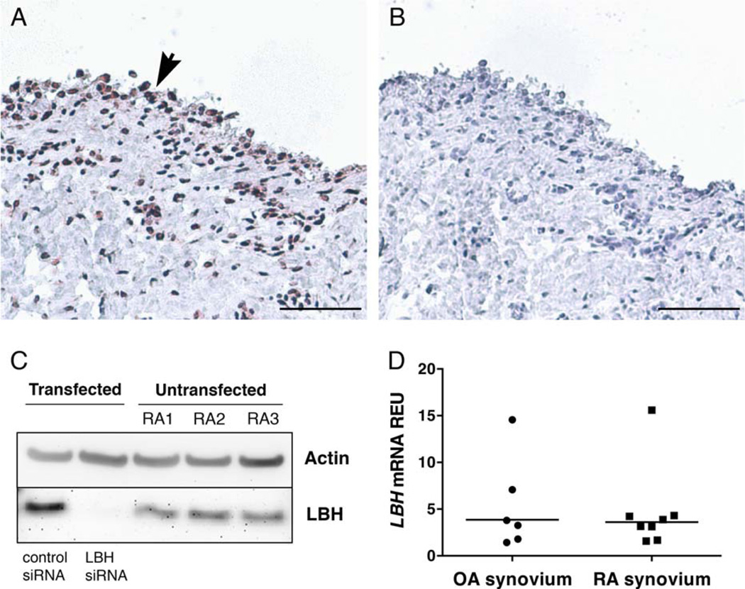
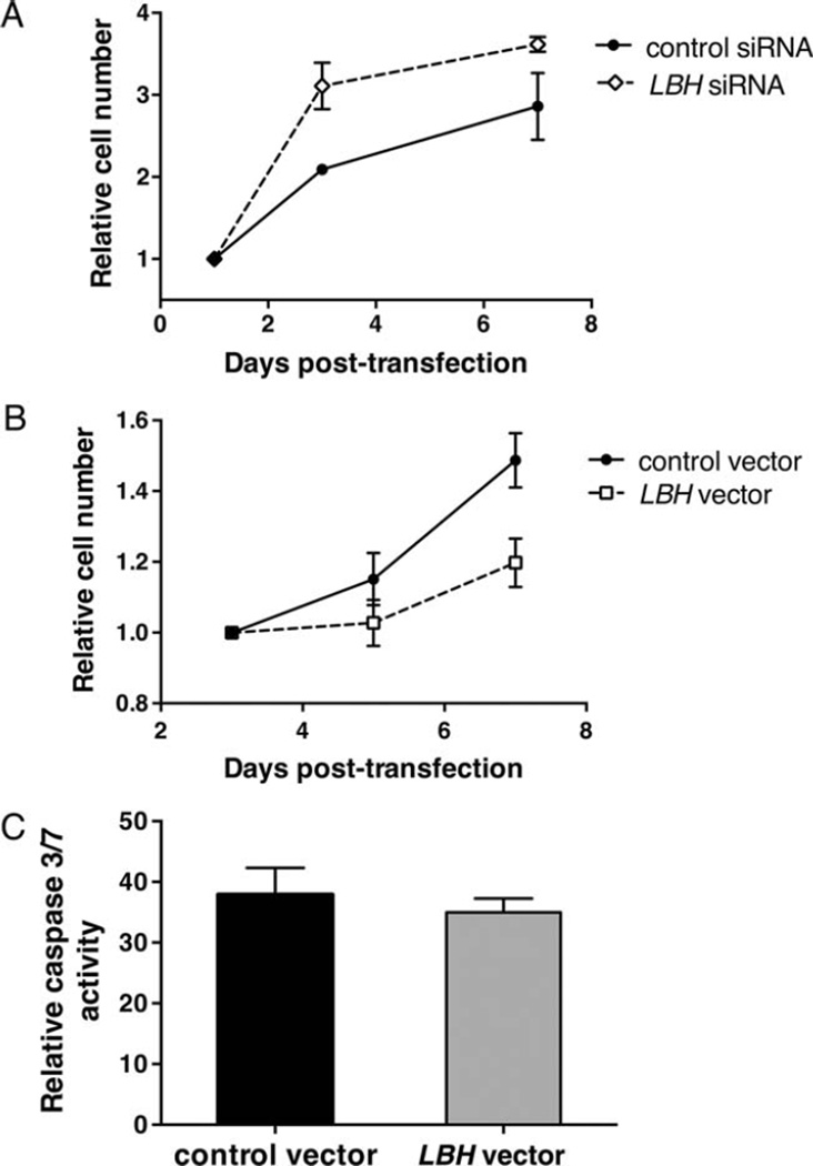
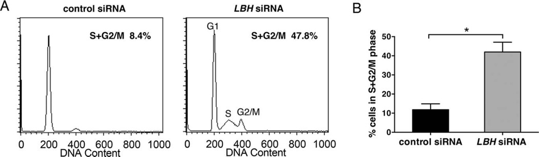
Results: LBH was expressed in the synovial lining layer in patients with RA. Transforming growth factor β1 significantly increased LBH expression in primary FLS, and platelet-derived growth factor BB decreased it. Pathway analysis of the transcriptome of LBH-deficient FLS compared to control FLS identified "cellular growth and proliferation" as the most significantly enriched pathway. In growth assays, LBH deficiency increased FLS proliferation. Conversely, LBH overexpression significantly inhibited cell growth. Cell cycle analysis demonstrated a marked increase in cells entering the cell cycle in LBH-deficient FLS compared to controls. LBH did not alter apoptosis.
Conclusion: LBH is a candidate gene for synovial pathology in RA. It is regulated by growth factors and modulates cell growth in primary FLS. Our data suggest a novel mechanism for synovial intimal hyperplasia and joint damage in RA.
© 2015, American College of Rheumatology.
Figures


Similar articles
-
The proto-oncogene survivin splice variant 2B is induced by PDGF and leads to cell proliferation in rheumatoid arthritis fibroblast-like synoviocytes.Sci Rep. 2015 May 22;5:9795. doi: 10.1038/srep09795. Sci Rep. 2015. PMID: 25997820 Free PMC article.
-
LBH Gene Transcription Regulation by the Interplay of an Enhancer Risk Allele and DNA Methylation in Rheumatoid Arthritis.Arthritis Rheumatol. 2016 Nov;68(11):2637-2645. doi: 10.1002/art.39746. Arthritis Rheumatol. 2016. PMID: 27159840 Free PMC article.
-
GREM1 Is a Key Regulator of Synoviocyte Hyperplasia and Invasiveness.J Rheumatol. 2016 Mar;43(3):474-85. doi: 10.3899/jrheum.150523. Epub 2016 Feb 1. J Rheumatol. 2016. PMID: 26834210
-
Synovial fibroblasts as potential drug targets in rheumatoid arthritis, where do we stand and where shall we go?Ann Rheum Dis. 2022 Jul 12;81(8):1055-1064. doi: 10.1136/annrheumdis-2021-222021. Ann Rheum Dis. 2022. PMID: 35715191 Free PMC article. Review.
-
Precision medicine in rheumatoid arthritis.Best Pract Res Clin Rheumatol. 2022 Mar;36(1):101742. doi: 10.1016/j.berh.2022.101742. Epub 2022 Mar 2. Best Pract Res Clin Rheumatol. 2022. PMID: 35248489 Free PMC article. Review.
Cited by
-
Novel Insights Into Rheumatoid Arthritis Through Characterization of Concordant Changes in DNA Methylation and Gene Expression in Synovial Biopsies of Patients With Differing Numbers of Swollen Joints.Front Immunol. 2021 Apr 22;12:651475. doi: 10.3389/fimmu.2021.651475. eCollection 2021. Front Immunol. 2021. PMID: 33968050 Free PMC article.
-
Construction of a ceRNA Network Related to Rheumatoid Arthritis.Genes (Basel). 2022 Apr 6;13(4):647. doi: 10.3390/genes13040647. Genes (Basel). 2022. PMID: 35456453 Free PMC article.
-
Fine-mapping cis-regulatory variants in diverse human populations.Elife. 2019 Jan 16;8:e39595. doi: 10.7554/eLife.39595. Elife. 2019. PMID: 30650056 Free PMC article.
-
Identification of Shared Genes Between Ischemic Stroke and Parkinson's Disease Using Genome-Wide Association Studies.Front Neurol. 2019 Mar 28;10:297. doi: 10.3389/fneur.2019.00297. eCollection 2019. Front Neurol. 2019. PMID: 30984102 Free PMC article.
-
Bacterial and viral pathogen-associated molecular patterns induce divergent early transcriptomic landscapes in a bovine macrophage cell line.BMC Genomics. 2019 Jan 8;20(1):15. doi: 10.1186/s12864-018-5411-5. BMC Genomics. 2019. PMID: 30621583 Free PMC article.
References
-
- Van Vollenhoven RF. Rheumatoid arthritis in 2012: progress in RA genetics, pathology and therapy. Nat Rev Rheumatol. 2013;9:70–72. - PubMed
Publication types
MeSH terms
Substances
Grants and funding
LinkOut - more resources
Full Text Sources
Other Literature Sources
Medical
Miscellaneous
