Case Reports
. 2015 May;77(5):840-50.
doi: 10.1002/ana.24389. Epub 2015 Mar 27.

Defective fast inactivation recovery of Nav 1.4 in congenital myasthenic syndrome

Affiliations
Case Reports

Defective fast inactivation recovery of Nav 1.4 in congenital myasthenic syndrome

W David Arnold et al. Ann Neurol. 2015 May.

Abstract

Objective: To describe the unique phenotype and genetic findings in a 57-year-old female with a rare form of congenital myasthenic syndrome (CMS) associated with longstanding muscle fatigability, and to investigate the underlying pathophysiology.

Methods: We used whole-cell voltage clamping to compare the biophysical parameters of wild-type and Arg1457His-mutant Nav 1.4.

Results: Clinical and neurophysiological evaluation revealed features consistent with CMS. Sequencing of candidate genes indicated no abnormalities. However, analysis of SCN4A, the gene encoding the skeletal muscle sodium channel Nav 1.4, revealed a homozygous mutation predicting an arginine-to-histidine substitution at position 1457 (Arg1457His), which maps to the channel's voltage sensor, specifically D4/S4. Whole-cell patch clamp studies revealed that the mutant required longer hyperpolarization to recover from fast inactivation, which produced a profound use-dependent current attenuation not seen in the wild type. The mutant channel also had a marked hyperpolarizing shift in its voltage dependence of inactivation as well as slowed inactivation kinetics.

Interpretation: We conclude that Arg1457His compromises muscle fiber excitability. The mutant fast-inactivates with significantly less depolarization, and it recovers only after extended hyperpolarization. The resulting enhancement in its use dependence reduces channel availability, which explains the patient's muscle fatigability. Arg1457His offers molecular insight into a rare form of CMS precipitated by sodium channel inactivation defects. Given this channel's involvement in other muscle disorders such as paramyotonia congenita and hyperkalemic periodic paralysis, our study exemplifies how variations within the same gene can give rise to multiple distinct dysfunctions and phenotypes, revealing residues important in basic channel function.


Conflict of interest statement

Potential Conflicts of Interest

Nothing to report.

Figures

FIGURE 1
FIGURE 1
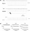
Electromyographic findings: (Left) 3Hz repetitive nerve stimulation (RNS) with no evidence of compound muscle action potential (CMAP) decrement; (Middle and Right) 10Hz RNS with CMAP decrement (−8.2% from 1st to 4th CMAP, and −17.6% from 1st to 10th CMAP) and 20Hz RNS with CMAP decrement (−4% from 1st to 4th CMAP, and −18.4% from 1st to 10th CMAP), respectively.
FIGURE 2
FIGURE 2
Chromatographs and pedigree. The pedigree and corresponding chromatograms illustrate the segregation of c.4370G>A, p.Arg1457His (R1457H) in the reported family (vertical arrows). The proband, who is homozygous for c.4370G>A and the only affected member of the family, is fully shaded (diagonal arrow), whereas the rest of the family members, who are heterozygous for c.4370G>A, are half shaded. [Color figure can be viewed in the online issue, which is available at www.annalsofneurology.org.]
FIGURE 3
FIGURE 3
Topology of Nav1.4 and location of Arg1457. The Nav1.4 protein comprises 1,836 amino acid residues falling into 4 homologous domains (D1–D4) of 6 transmembrane regions each (S1–S6). In a pseudotetramer, D1 through D4 fold around a central aqueous pore that mediates all ion flow. Between S5 and S6 of each domain is a largely hydrophobic membrane-reentrant loop known as the pore loop (“P-loop”) that forms the outer vestibule of the pore and includes conserved charged residues that confer selectivity for sodium. Voltage sensitivity of the channel is provided by positively charged amino acids (arginines and lysines, gray background) in the S4 regions, which move in the electric field set by the membrane potential. Both the N-terminus and C-terminus map to the cell’s interior. The location of the Arg1457His exchange is shown as a black dot in D4/S4, which is enlarged at the bottom of the figure, along with an alignment against all human Nav isoforms (National Center for Biotechnology Information identifiers provided in parentheses). The numbers in the center provide the position of the N-terminal residue in D4/S4 for each isoform. Arginine 1457 is emphasized in black; it is conserved across all human Nav channels.
FIGURE 4
FIGURE 4
Biophysical characterization of Nav1.4 Arg1457His based on heterologous whole-cell currents. (A) Normalized current– voltage relationship for wild-type (solid squares) and Arg1457His (open squares) channels. Note the small but discernible shift in the voltage triggering the maximal response. (B) Voltage dependence of activation calculated from the data shown in A. Arg1457His channels show a subtle depolarizing shift in half-maximal activation. (C) Steady-state fast inactivation. The curves for wild-type and Arg1457His channels clearly separate, as the mutant fast-inactivates much more readily. At potentials where the wild type is mostly unaffected (eg, −100 and −90 mV), Arg1457His shows a marked reduction in availability. (D) Recovery from fast inactivation. Arg1457His channels show a unique recovery profile. As in wild-type channels, biexponential fitting adequately reproduced the experimental mutant data, which could be explained by 2 distinct channel populations or by 2 separate gating modes, recovering quickly (τ1) or more slowly (τ2). The former dominates in the wild type (88%) but not in Arg1457His (49%). What is more, τ1 is much larger in Arg1457His (25.1 ± 5.6 milliseconds) than in the wild-type channels (2.7 ± 0.3 milliseconds). (E) Voltage-dependent fast inactivation kinetics. Displayed are the averaged time constants following single exponential fitting of the Na+-transient decay. All fitting data are listed in Tables 1 and 2.
FIGURE 5
FIGURE 5
Enhanced use dependence in Arg1457His channels. (A) HEK cells expressing either wild-type or R1457H-mutant Nav1.4 were clamped in the whole-cell configuration and held at −120mV to be repeatedly stepped to −10mV (5 milliseconds, 30Hz). The dotted line demarks the current amplitude of the first sodium transient. Note how wild-type channels remain unaffected by the stimulation paradigm, whereas the mutant channels show a significant current reduction with the second pulse (arrow). The traces were normalized to each other based on the first transient’s amplitude. (B) Averaged use dependence at the indicated stimulation frequencies. The maximal current response for each pulse (Pn) was normalized to the amplitude encountered in the first pulse (P1) for wild-type (solid squares, n = 5) or R1457H-mutant (open squares, n = 5). Note that P1,n/P1 is equal to unity for all data sets. For clarity, only every second (10 and 77Hz) or sixth value (30Hz) is displayed.
FIGURE 6
FIGURE 6
Slow inactivation analyses. HEK cells expressing wild-type (solid squares) or R1457H-mutant (open squares) Nav1.4 were held at −120mV and subjected to the pulse protocols similar to Figure 4C and D, with the difference that the duration of the first pulse (P1) was extended to 10 seconds. (A) Onset of slow inactivation as assessed by increasingly long pulses to −10mV (P1), brief fast inactivation recovery, and a second −10mV test pulse (P2). Wild-type, n = 13; R1457H-mutant, n = 11. (B) Steady-state slow inactivation. Wild-type, n = 16; R1457H-mutant, n = 5. (C) Recovery from slow inactivation. Wild-type, n=12; R1457H-mutant, n = 9. In all experiments, 200 milliseconds at −120mV just prior to the test pulse (P2) ensured full recovery from fast inactivation (see Fig 4D). The data are displayed as mean ± standard error of the mean. Further details are provided in the Case Report section.

References

    1. Engel AG. Current status of the congenital myasthenic syndromes. Neuromuscul Disord. 2012;22:99–111. - PMC - PubMed
    1. Beeson D. Synaptic dysfunction in congenital myasthenic syndromes. Ann N Y Acad Sci. 2012;1275:63–69. - PubMed
    1. Engel AG, Shen XM, Selcen D, Sine SM. What have we learned from the congenital myasthenic syndromes. J Mol Neurosci. 2010;40:143–153. - PMC - PubMed
    1. Slater CR. Structural factors influencing the efficacy of neuromuscular transmission. Ann N Y Acad Sci. 2008;1132:1–12. - PubMed
    1. Ruff RL, Lennon VA. End-plate voltage-gated sodium channels are lost in clinical and experimental myasthenia gravis. Ann Neurol. 1998;43:370–379. - PubMed

Publication types

Substances