. 2015 Feb 26;518(7540):507-11.
doi: 10.1038/nature14174. Epub 2015 Feb 18.

Crystal structure of the V(D)J recombinase RAG1-RAG2

Affiliations

Crystal structure of the V(D)J recombinase RAG1-RAG2

Min-Sung Kim et al. Nature. .

Abstract

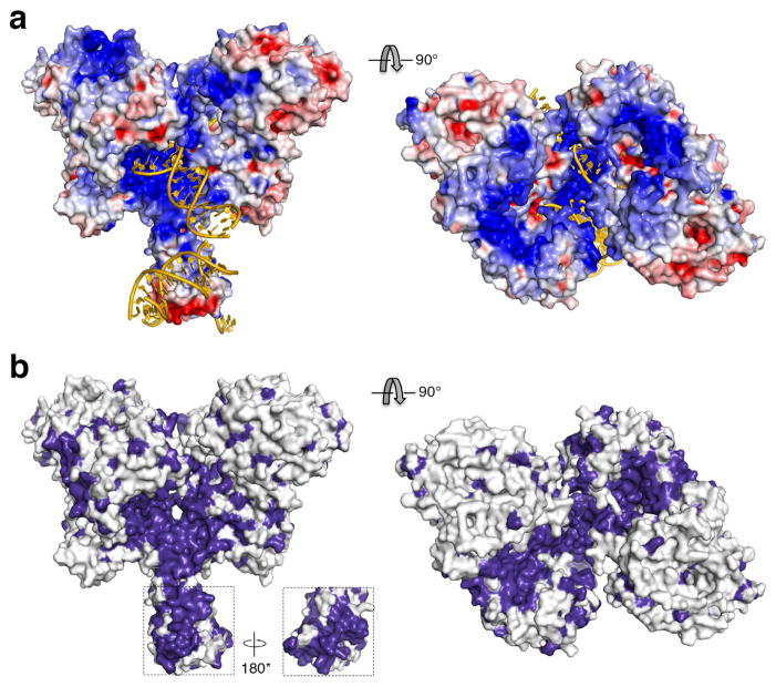
V(D)J recombination in the vertebrate immune system generates a highly diverse population of immunoglobulins and T-cell receptors by combinatorial joining of segments of coding DNA. The RAG1-RAG2 protein complex initiates this site-specific recombination by cutting DNA at specific sites flanking the coding segments. Here we report the crystal structure of the mouse RAG1-RAG2 complex at 3.2 Å resolution. The 230-kilodalton RAG1-RAG2 heterotetramer is 'Y-shaped', with the amino-terminal domains of the two RAG1 chains forming an intertwined stalk. Each RAG1-RAG2 heterodimer composes one arm of the 'Y', with the active site in the middle and RAG2 at its tip. The RAG1-RAG2 structure rationalizes more than 60 mutations identified in immunodeficient patients, as well as a large body of genetic and biochemical data. The architectural similarity between RAG1 and the hairpin-forming transposases Hermes and Tn5 suggests the evolutionary conservation of these DNA rearrangements.


Conflict of interest statement

The authors declare no competing financial interests.

Figures

Extended Data Figure 1
Extended Data Figure 1. Structure determination
(a) The plot of correlation coefficient (CC) of anomalous signal versus resolution. The red line indicates the cutoff of CC=0.3. Merging data from the two best crystals produced a better CC than merging data from all six crystals. The data processing procedure is outlined above the plot . (b) The SAD experimental map contoured at 1.3σ showed the content of an asymmetric unit. The anomalous peaks of selenium atoms are shown in red. (c) A typical crystal of RAG1/2. (d) The content of crystals was examined by protein and DNA denaturing gels after a thorough wash of the crystals and stained by Coomassie Blue and SYBR-Green. To confirm the 1:1 molar ratio of 12 and 23RSS DNA, 32P-labeled input RSS DNAs and those in SEC complexes before and after crystallization are shown beneath the SYBR-Green stained DNA gel. (e) Transposition assay of the purified SEC (RAG1/2-12/23RSS DNA complex) used for crystallization. Supercoiled pUC19 (sc, with a small amount of open circle, oc) was the target; it was linearized by HindIII as a control. The SEC (0.25, 0.5 and 1.0 μM) was active in concerted transposition and thus linearizing pUC19. In contrast, RAG1/2 or HMGB1 (0..5μM) each alone was not active. (f) Crystal packing of neighboring RAG1/2 complexes (shown in dark and light colors) occludes one nonamer-binding site in each heterotetramer of RAG1/2.
Extended Data Figure 2
Extended Data Figure 2. RAG2 core fragment (1–351aa) is active
(a) Sequence alignment of RAG2 from mouse (320–387aa), human, rat and Xenopus with predicted secondary structures shown above. (b) Core RAG2 (1–387) and two further truncated RAG2 variants (1–351 and 1–367) were constructed with a non-cleavable N-terminal MBP tag and co-expressed with the tag-less core RAG1. The Coomassie Blue R-250 stained SDS gel shows the purified RAG1/RAG2 complexes. (c) Purified RAG1/2 complexes with truncated RAG2 variants are equally active in cleaving a 32P-labeled 12RSS DNA (in the presence of a 23RSS and Mg2+, as examined by TBE-Urea gel). (d) Elution profiles of RAG1/RAG2 (both long and short forms) complexed with DNA from Superdex-200 (S200) in a low salt buffer (50mM HEPES 7.0, 60 mM KCl, 1mM maltose and 2 mM DTT). Regardless of the length of RAG2, the major S200 eluant peak came out at the same time point and contained RAG1, RAG2 (1–351 or 1–387) and HMGB1 proteins, as shown in the SDS gel (right insert), as well as 12 and 23RSS oligonucleotides, as confirmed by a TBE-Urea gel stained by SYBR-Green (left insert).
Extended Data Figure 3
Extended Data Figure 3. Comparison of RAG2 with β-propeller and β-pinwheel structures
KLHL2 (PDB: 4CHB) is selected to represent the β-propeller proteins, and the C-terminal domain (CTD) of GyrA (PDB: 1SUU) is selected to represent the β-pinwheel structures. After superposition, RAG2, KLHL2 and GyrA are shown side-by-side individually in two orthogonal views. Each structure is colored from N- to C-terminus in blue to red rainbow colors. The loops in RAG2 that interact with RAG1 are labeled. The six β blades are named by Roman numerals, I to VI from N to C terminus; four β strands in each blade are named by Arabic numerals, 1 to 4.
Extended Data Figure 4
Extended Data Figure 4. Comparison of RAG1 and NBD-DNA complex
(a) The NBD in the RAG1/2 core complex (blue and green) superimposes well with the published structure of the NBD-DNA complex (PDB: 3GNA, protein colored yellow) . (b) The twelve SCID/OS mutations in the NBD domain are mapped onto the crystal structure of the NBD-DNA complex. Six SCID/OS (R391 to R407) mutations are located on a positively charged surface patch that interacts with the nonamer; five remaining SCID/OS mutations (L408 to A441) appear to affect the structural integrity of the NBD, and R446 may interact with the spacer DNA in each RSS.
Extended Data Figure 5
Extended Data Figure 5. Transposases that form a hairpin intermediate
(a) Hermes (PDB: 4D1Q), (b) bacterial Tn5 (PDB: 1MUS), and (c) RAG1 dimers are shown as ribbon diagrams in two orthogonal views, with the dyad perpendicular to the viewing plane (left) or in the plane (right). Each dimer consists of a cyan and a green subunit. The catalytic RNH domains are highlighted in pink, and the conserved catalytic residues are shown as red ball-and-sticks. The catalytic divalent metal ions are shown as green spheres if present. The DNAs, colored in yellow (cleaved by the cyan subunit) and orange (cleaved by the green subunit), have similar orientations in the Hermes and Tn5 complexes (as indicated by the arrows). Arrows with dashed outlines indicate that the DNAs are in the back of the viewing plane. Notably, the pair of RNH domains is oriented similarly in all three cases. The predicted orientations of DNAs bound to RAG1 are indicated by the yellow and orange arrows, and the α-helices connected to the third catalytic carboxylates (shown in light purple) probably bridge two DNAs in RAG1 recombinase as in Hermes and Tn5.
Extended Data Figure 6
Extended Data Figure 6. Transposases that do not form a hairpin intermediate
(a) Retroviral integrase from Prototype foamy virus (Pfv, PDB: 3OS0), (b) bacterial MuA transposase (PDB: 4FCY), and (c) eukaryotic Mos1 mariner transposase (PDB: 3HOT) are shown in comparable views and same representations as Hermes, Tn5 and RAG1/2 in Extended Data Figure 5. Each catalytic dimer consists of a cyan and a green subunit. Two accessory subunits in Pfv are shown in light blue and green, and two accessory subunits of the MuA structure are omitted for clarity. The catalytic RNH domains are highlighted in pink. The DNAs, colored in yellow (cleaved by the cyan subunit) and orange (cleaved by the green subunit), have similar orientations (within 30°) as indicated by the arrowheads, but each differs more than 90° from the corresponding DNA in Hermes or Tn5 transposase. The gray DNA in the MuA complex represents the target of transposition. Among these three recombinases, the α-helix that follows the third catalytic carboxylate (colored in light purple) does not cross over to interact with a second DNA.
Extended Data Figure 7
Extended Data Figure 7. Surface potential and conservation of RAG1/2 complex
(a) Orthogonal views of the electrostatic potential surface of the RAG1/2 structure. Blue indicates positive charges, and red negative. (b) Orthogonal views of the molecular surface of RAG1/2 with absolutely conserved residues highlighted in deep purple. The NBD is well conserved. The views with dyad in the plane here are related to the image shown in Fig. 5c by ~50° rotation around the dyad.
Fig. 1
Fig. 1
Structure determination of RAG1/2 recombinase. (a) Procedure of assembling SEC from purified RAG1/2, RSS DNAs and HMGB1. The MBP tags were cleaved off by PreScission protease after SEC formation. (b) The RAG1/2 – DNA complex was purified away from MBP tags, free DNA, and HMGB1 on a Superdex-200 column. The eluted peak contains RAG1/2 protein and RSS DNAs, as shown in the protein and DNA denaturing gels stained by Coomassie Blue and SYBR-Green, respectively. (c, d) The experimental electron density map calculated from merging (c) two best SAD datasets or (d) all six SAD datasets.
Fig. 2
Fig. 2
Crystal structure of RAG1/2. (a) The front and (b) top view of the RAG1/2 heterotetramer. The two RAG1 chains are shown in blue and green ribbon diagrams, and both RAG2 subunits are shown in magenta. The active sites are highlighted by the three carboxylates shown as red sticks. The zinc ions are shown as dark red spheres. The distance between the two active sites is ~45Å (marked by the red double arrowheads). The disordered loop of residues 608–614 in RAG1 near the RAG1/2 interface is marked by a dotted line and grey arrowhead.
Fig. 3
Fig. 3
The RAG1 structure. (a) A diagram of RAG1 domains with boundaries indicated by residue numbers. (b) Cartoons of RAG1 dimer. One subunit is color-coded as in (a), and the other in silver except for the preR and ZnC2 domains. (c) An orthogonal view of (b). (d) The Zn2+ coordination by two Cys (of ZnC2) and two His residues (of ZnH2). (e) The DDBD and CTD domains. (f) The RNH and a portion of preR domain with the catalytic carboxylates shown in red sticks. In panels b, d e, f, SCID/OS mutations are shown as colored sticks with black labels.
Fig. 4
Fig. 4
The interface between RAG1 and RAG2. (a) One side of the doughnut-shaped RAG2 interacts with the preR, RNH and ZnC2 domains of RAG1 (color-coded as in Fig. 3). The SCID/OS mutations in RAG2 are shown and labeled in black. (b) An orthogonal view of the RAG1/2 interface. The Kelch repeats of RAG2 are labeled I to VI. (c) Based on (b), the interface of RAG1 and RAG2 is shown as an open book. The regions at the interface are indicated according to the color code, and SCID/OS mutations are labeled in black. The mirrored arrowheads and boxes indicate matching surfaces.
Fig. 5
Fig. 5
A RAG1/2-DNA model. (a) Superposition of the RNH domains of RAG1 (red active site) and Hermes (PDB: 4D1Q ). (b) The superposition places the 16bp DNA of the Hermes-DNA complex in the RAG1 active site. The DNA is colored yellow (the first 7 bp) and gold. The second DNA is shown without additional manipulation. The α-helices bridging the two DNAs are colored lilac. (c) The RAG1/2-RSS DNA model resulting from superposition with the Hermes and the NBD–DNA complex (12bp, PDB: 3GNA) . (d) DNA cleavage by hairpin-forming bacterial and eukaryotic transposases. The recognition sequences are represented by orange triangles.

References

    1. Sakano H, Huppi K, Heinrich G, Tonegawa S. Sequences at the somatic recombination sites of immunoglobulin light-chain genes. Nature. 1979;280:288–294. - PubMed
    1. Lewis SM. The mechanism of V(D)J joining: lessons from molecular, immunological, and comparative analyses. Adv Immunol. 1994;56:27–150. - PubMed
    1. Gellert M. V(D)J recombination: RAG proteins, repair factors, and regulation. Annu Rev Biochem. 2002;71:101–132. doi: 10.1146/annurev.biochem.71.090501.150203090501.150203. [pii] - DOI - PubMed
    1. Schatz DG, Swanson PC. V(D)J recombination: mechanisms of initiation. Annu Rev Genet. 2011;45:167–202. doi: 10.1146/annurev-genet-110410-132552. - DOI - PubMed
    1. Deriano L, Roth DB. Modernizing the nonhomologous end-joining repertoire: alternative and classical NHEJ share the stage. Annu Rev Genet. 2013;47:433–455. doi: 10.1146/annurev-genet-110711-155540. - DOI - PubMed

Publication types

MeSH terms

Associated data