. 2015 Mar 12;519(7542):199-202.
doi: 10.1038/nature14245. Epub 2015 Feb 18.

Cas9 specifies functional viral targets during CRISPR-Cas adaptation

Affiliations

Cas9 specifies functional viral targets during CRISPR-Cas adaptation

Robert Heler et al. Nature. .

Abstract

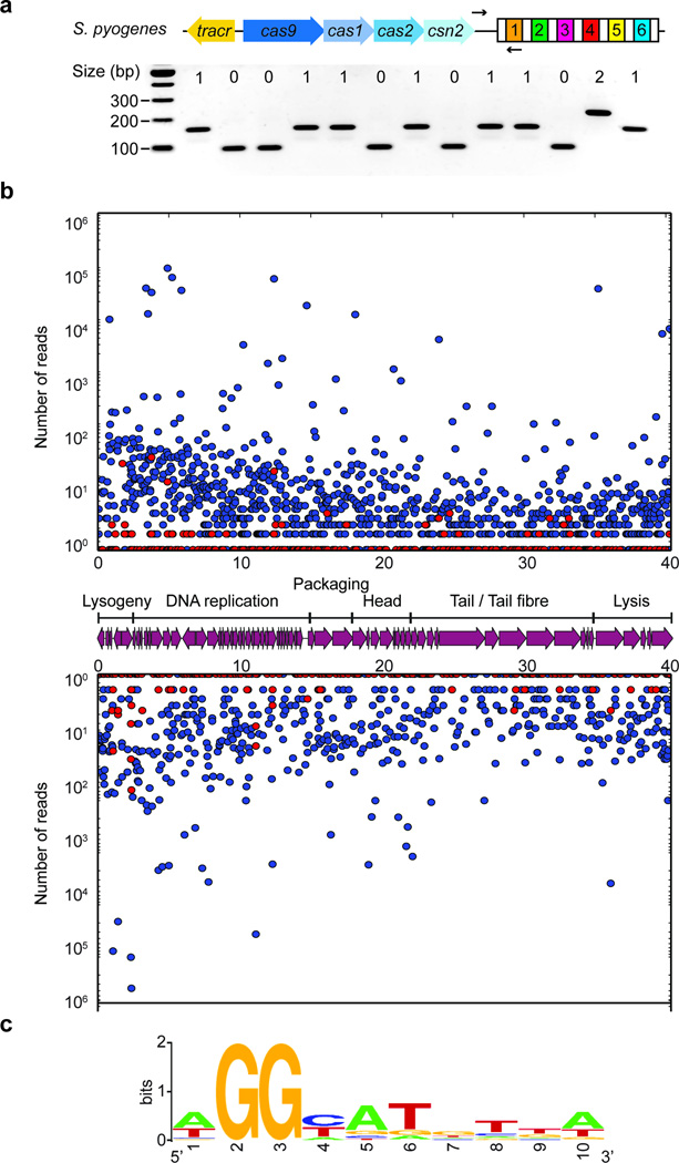
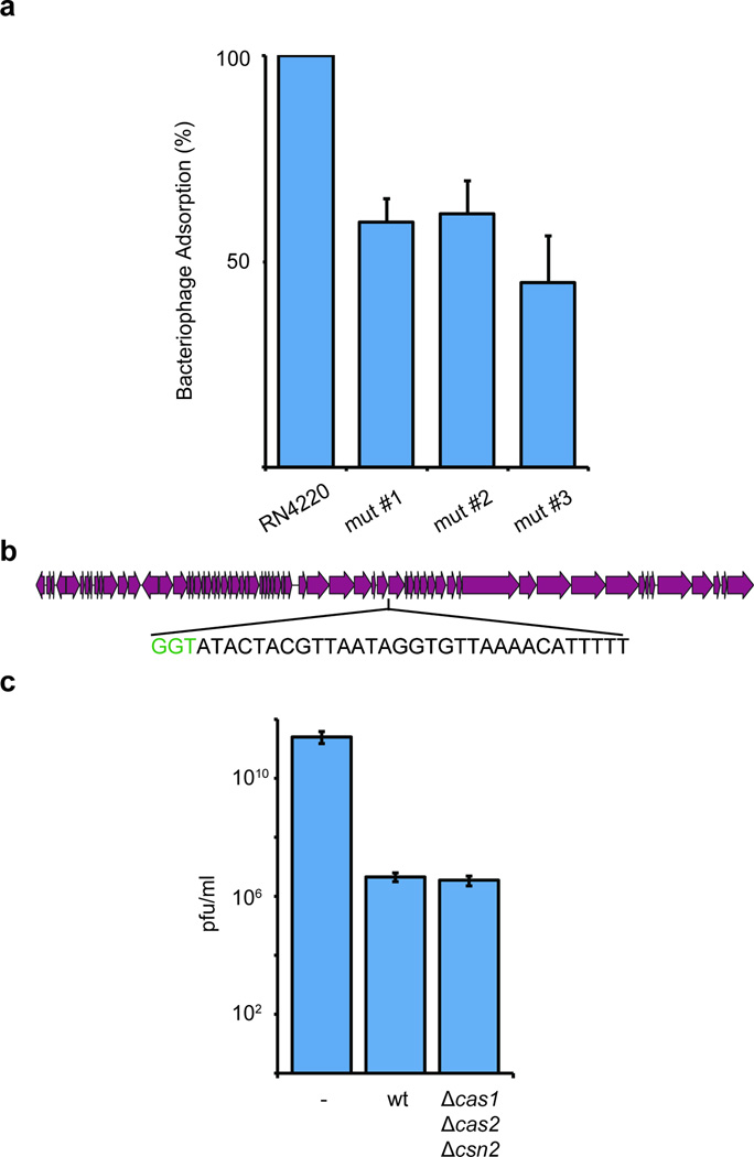
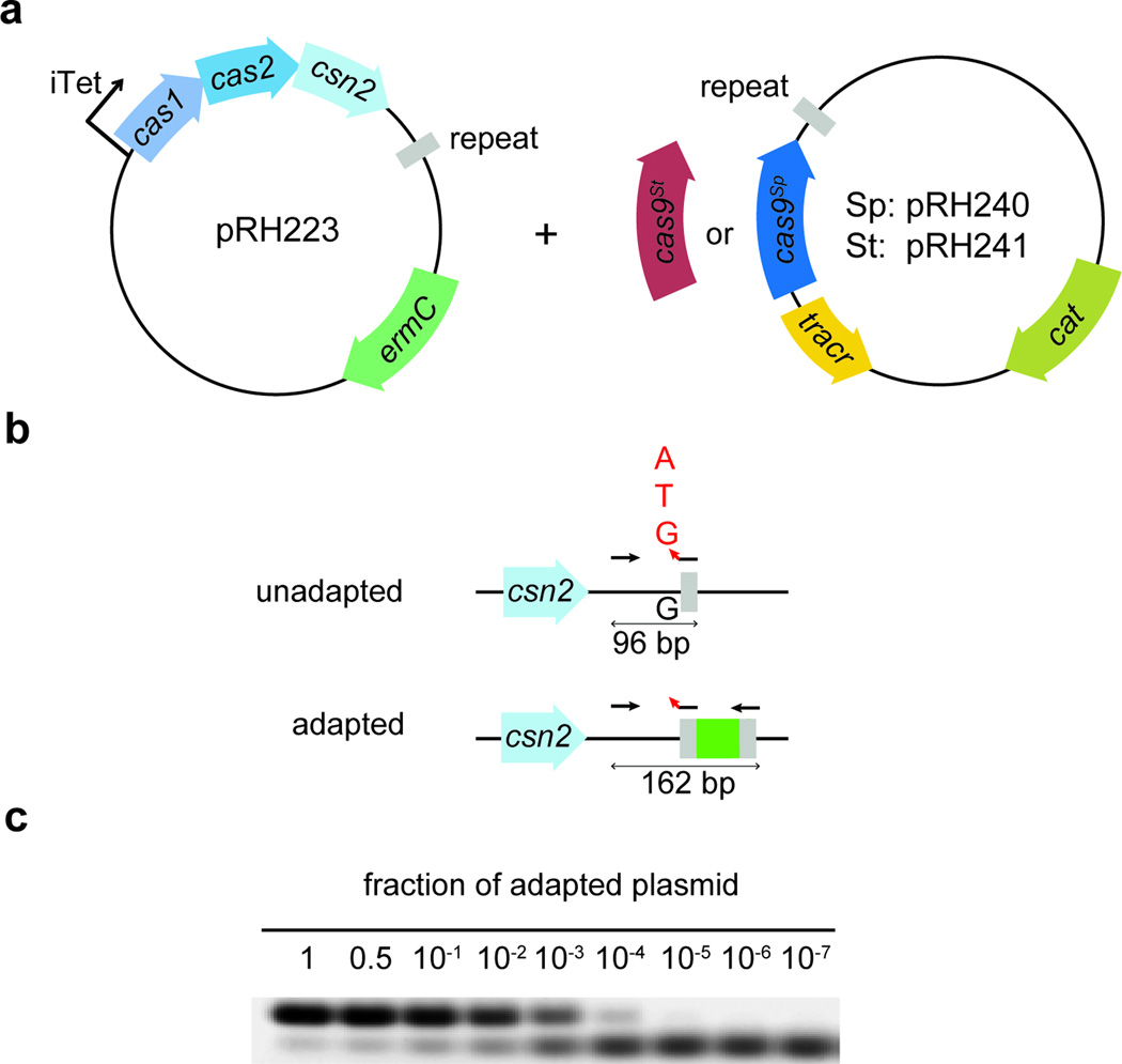
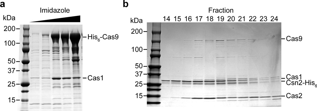
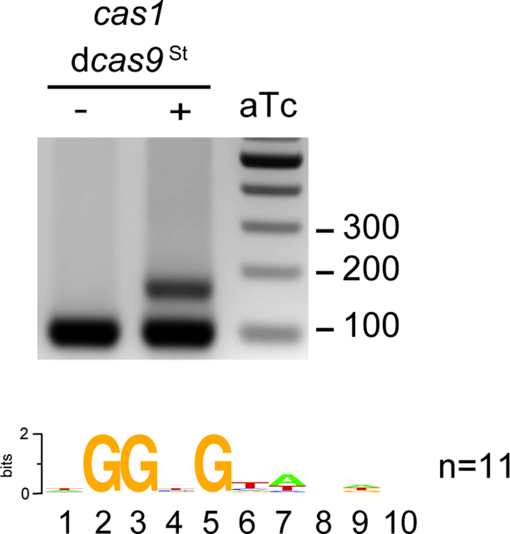
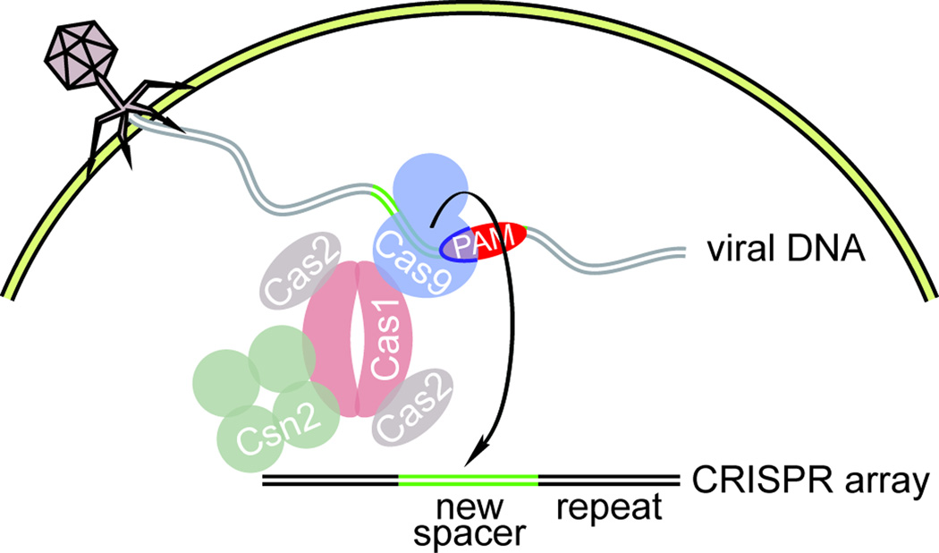
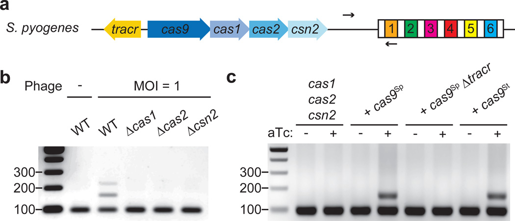
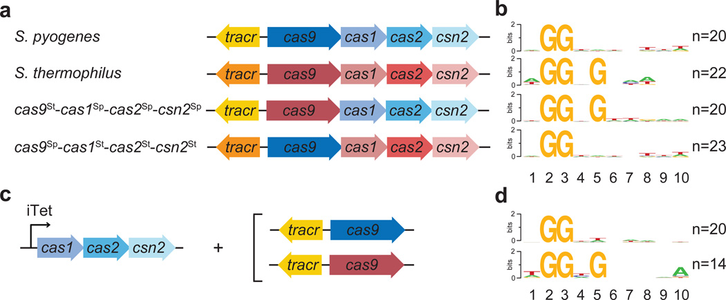
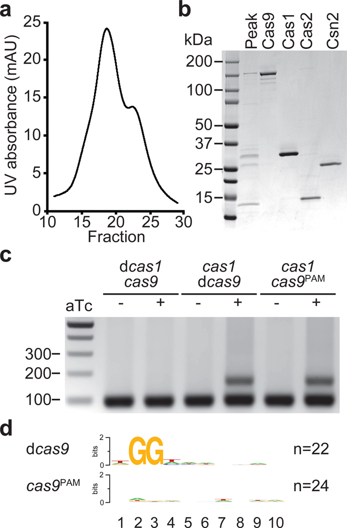
Clustered regularly interspaced short palindromic repeat (CRISPR) loci and their associated (Cas) proteins provide adaptive immunity against viral infection in prokaryotes. Upon infection, short phage sequences known as spacers integrate between CRISPR repeats and are transcribed into small RNA molecules that guide the Cas9 nuclease to the viral targets (protospacers). Streptococcus pyogenes Cas9 cleavage of the viral genome requires the presence of a 5'-NGG-3' protospacer adjacent motif (PAM) sequence immediately downstream of the viral target. It is not known whether and how viral sequences flanked by the correct PAM are chosen as new spacers. Here we show that Cas9 selects functional spacers by recognizing their PAM during spacer acquisition. The replacement of cas9 with alleles that lack the PAM recognition motif or recognize an NGGNG PAM eliminated or changed PAM specificity during spacer acquisition, respectively. Cas9 associates with other proteins of the acquisition machinery (Cas1, Cas2 and Csn2), presumably to provide PAM-specificity to this process. These results establish a new function for Cas9 in the genesis of prokaryotic immunological memory.


Conflict of interest statement

The authors have no conflicting financial interests.

Figures

Extended Data Figure 1
Extended Data Figure 1. The S. pyogenes type II CRISPR-Cas system displays a strong bias for the acquisition of spacers matching viral protospacers with NGG PAMs
a, Analysis of bacteriophage-insensitive mutant colonies using PCR and agarose gel electrophoresis, representative of five technical replicates. Bacteria and phage were mixed in top agar and incubated overnight. DNA was isolated from individual colonies resistant to phage infection and used as template for a PCR reaction with primers (arrows) H182 and H183 (Extended Data Table 2), which amplify the 5’ end of the S. pyogenes CRISPR array. The size of the PCR band indicates the number of new spacers (shown at the top of the gel). Cells without additional spacers resist infection by a CRISPR-independent mechanisms, presumably envelope resistance. b, Analysis of acquired spacers during phage infection of a population of bacteria carrying the S. pyogenes type II CRISPR-Cas system. Liquid cultures of bacteria were infected with phage, surviving cells were collected at the end of the infection, DNA extracted and used as template for a PCR reaction as described above. Amplification products were separated by agarose gel electrophoresis and the DNA of the bands corresponding to products with additional spacers was extracted and sent for Mi-Seq next generation sequencing. Reads corresponding to newly acquired spacers were plotted according to their position in the phage ϕNM4γ4 genome (x-axis) and their abundance (y-axis). Each dot represents a unique spacer sequence; blue and red dots indicate a corresponding protospacer with an NGG or non-NGG PAM. Top and bottom plots indicate protospacers in the top and bottom strands of the ϕNM4γ4 DNA. The map as well as the different functions of the phage genes are indicated in between the plots. The raw data used to make this graph is in the Supplementary file. c, Weblogo showing the conservation of the 5’ flanking sequences of 10,000 protospacers randomly selected from the experiment shown in b. Absolute conservation of the NGG PAM was observed.
Extended Data Figure 2
a, Analysis of bacteriophage-resistant mutants that do not acquire a new spacer. Three colonies that survived phage infection in our in-plate adaptation assay (Fig. S1a) were subjected to phage adsorption assay. Briefly, surviving colonies as well as the wild-type S. aureus RN4220 control were grown in liquid and mixed with bacteriophage. After a brief incubation, cells were pelleted by centrifugation and the phages present in the supernatant (unable to bind and infect cells) were counted on a lawn of sensitive cells. The number of plaque-forming units (pfu) of a control experiment in the absence of host cells were used to determine the 100% free-phage, or 0% adsorption, value. No plaques were observed in the control experiment using wild-type cells and this value was used to set the 100% adsorption limit. The three CRISPR-independent, bacteriophage-resistant mutants displayed a marked defect in phage adsorption (about 50 %), indicating that most likely they carry envelope resistance mutations. Error bars: mean ± s.d. (n=3). b, cas1, cas2 and csn2 are not required for the execution of immunity using previously acquired spacers. Position within the phage ϕNM4γ4 genome of the type II CRISPR-Cas target used in this experiment. The protospacer sequence is in the bottom strand (shown in 3’–5’ direction) and flanked by a TGG PAM (in green). c, Comparison of immunity provided by a type II CRISPR-Cas system programmed to target the sequence shown in panel a in the presence (wild-type, wt) or absence (Δcas1cas2, Δcsn2) of cas1, cas2 and csn2. Immunity is measured as the plaque forming units (pfu) of a φNM4γ4 phage lysate spotted on top agar lawns of S. aureus RN4220 cells containing no CRISPR system (−), a wild type S. pyogenes CRISPR-Cas type II system (wt, pRH233), or the same CRISPR-Cas systems with a deletion of cas1, cas2 and csn2 genes (Δcas1cas2, Δcsn2, pRH079). Error bars: mean ± s.d. (n=3).
Extended Data Figure 3
Extended Data Figure 3. Generation of an experimental system for the overexpression of cas1, cas2 and csn2 and the detection of spacer acquisition in the absence of phage infection
a, Plasmids used in the spacer acquisition experiments presented in Fig. 1c and Fig. 2c–d. pRH223 contains cas1, cas2 and csn2 from S. pyogenes under a tetracycline-inducible promoter. Cells containing this plasmid only acquired spacers when a second plasmid expressing cas9 was introduced, pRH240 or pRH241, containing the tracrRNA gene, the leader and first repeat from the S. pyogenes type II CRISPR-Cas system as well as cas9 from S. pyogenes (cas9Sp) or S. thermophilus (cas9St), respectively. The leader is a short, AT rich sequence immediately upstream of the first repeat that contains the promoter for the transcription of the CRISPR array. b, Highly sensitive PCR assay to enrich for amplification products of adapted CRISPR loci. Arrows indicate primer annealing position and direction. The forward primer (JW8) anneals on the leader. For the reverse primer, a cocktail of JW3, JW4 and JW5 was used. The three reverse primers anneal on the repeat and differ only in their 3’-end nucleotide that never matches the last nucleotide of the leader (red arrowhead). Because this nucleotide is critical for the annealing of the primers, loci that acquire spacers ending in A, C or T are preferentially amplified over unadapted loci. c, To quantify the sensibility of this technique, we mixed pGG32 (one repeat, unadapted) with pRH087 (repeat-spacer-repeat, adapted) in known ratios. The amplification of adapted plasmid was detected even when it represented 0.01% (104) of the total plasmid template, representative of three technical replicates. This highly sensitive PCR assay is not required to detect acquisition during phage infection, as in this case adapted cells survive and are enriched within the population, making their detection much easier.
Extended Data Figure 4
Extended Data Figure 4. Purification of a Cas9-Cas1-Cas2-Csn2 complexes
a, The cas9-cas1-cas2-csn2 operon of S. pyogenes SF370 was cloned into the pET16b vector (generating pKW07) to add an N-terminal histidyl tag to Cas9 and express all proteins in E. coli. Purification was performed using Ni-NTA affinity chromatography. SDS-PAGE followed by Coomassie stain of the purified proteins revealed a co-purifying protein that was identified as Cas1 by mass spectrometry, representative of five technical replicates. Mass spectrometry identification of all the eluted proteins co-purifying with Cas9 is shown in Extended Data Table 2. b, The cas9-cas1-cas2-csn2 operon of S. pyogenes SF370 was cloned into the pET23a vector (generating pKW06) to add an C-terminal histidyl tag to Csn2 and express all proteins in E. coli. Purification was performed using Ni-NTA affinity chromatography followed by ion exchange chromatography. The elution fractions that constituted the peak containing the complex (Fig. 3a) were separated by SDS-PAGE and visualized by Coomassie staining, representative of three technical replicates.
Extended Data Figure 5
Extended Data Figure 5. dCas9St can also support spacer acquisition
A plasmid derived from pRH241 containing mutations in the active site of St Cas9 (D10A, H847A; dCas9St) was used to characterize spacer acquisition in the absence of phage infection. Upon over-expression of Cas1, Cas2 and Csn2 using anydrotetracycline (aTc), we were able to detect spacer acquisition. Sequencing of spacers and alignment of the protospacer flanking sequences demonstrated the selection of an NGGNG PAM. Image is representative of three technical replicates.
Extended Data Figure 6
Extended Data Figure 6. A model for the selection of PAM-flanking spacers by Cas9
After injection of the phage DNA an adaptation complex formed by Cas9, Cas1, Cas2 and Csn2 uses the Cas9 PAM binding domain to specify functional protospacers, i.e., that are followed by the correct PAM. It is not known how the protospacer sequence is extracted from the viral DNA to become a spacer. In the “cut and paste” model, a nuclease, possibly Cas1, cuts the viral DNA to generate the spacer. In the “copy and paste” model the protospacer sequence is copied first. Once loaded with the selected protospacer sequence, this complex promotes the integration of this sequence into the CRISPR array, thus becoming a new spacer. Previous studies demonstrated that Cas1 dimerizes and interacts with Cas2 (ref.); Csn2 has been determined to form a tetramer.
Figure 1
Figure 1. Cas9 is required for spacer acquisition
a, Organization of the S. pyogenes type II CRISPR-Cas locus. Arrows indicate the annealing position of the primers used to check for the expansion of the CRISPR array. b, PCR-based analysis of liquid cultures to check for the acquisition of new spacer sequences in the presence or the absence of phage ϕNM4γ4 infection. Wild-type (WT) as well as different cas mutants were analyzed. Image is representative of three technical replicates. MOI; multiplicity of infection. c, Cultures over-expressing Cas1, Cas2 and Csn2 under the control of a tetracycline-inducible promoter were analyzed using PCR for spacer acquisition in the absence of phage infection. The strain was complemented with plasmids carrying either St or Sp Cas9 (see Extended Data Fig. 3), in the last case with or without the tracrRNA gene (Δtracr). Image is representative of three technical replicates. aTc; anhydrotetracycline.
Figure 2
Figure 2. Cas9 determines the PAM sequence of acquired spacers
a, c, Genetic composition of the CRISPR-Cas loci tested for spacer during phage infection (a), or in the absence of infection, with the experimental set up shown in Fig. S4 (c). b, d, Sequence logos obtained after the alignment of the 3’ flanking sequences of the protospacers matched by the newly acquired spacers in panels a and c, respectively. Numbers indicate the positions of the flanking nucleotides downstream from the spacer. n; number of sequences used in each alignment.
Figure 3
Figure 3. Cas9Sp PAM recognition domain is required for the acquisition of spacers with an NGG PAM sequence
a, Separation of the Cas9-Cas1-Cas2-Csn2 complex by ion exchange chromatography. b, SDS-PAGE of fraction 19 (peak) from the complex elution shown in panel a, representative of five technical replicates. The four proteins of the complex were individually purified and run alongside the purified fraction to identify each protein in the complex. c, Spacer acquisition was tested as in Fig. 1c in the presence or absence of different Cas1 or Cas9 activities. Image is representative of eight technical replicates. dCas1, nuclease-dead Cas1 (E220A mutation); dCas9, nuclease-dead Cas9 (D10A, H840A mutations); Cas9PAM lacks the PAM recognition function (R1333Q, R1335Q mutations). d, Sequence logos obtained after the alignment of the 3’ flanking sequences of the protospacers matched by the newly acquired spacers in panel c. Numbers indicate the positions of the flanking nucleotides downstream from the spacer. n; number of sequences used in each alignment.

Comment in

References

    1. Barrangou R, Marraffini LA. CRISPR-Cas systems: prokaryotes upgrade to adaptive immunity. Mol. Cell. 2014;54:234–244. - PMC - PubMed
    1. Barrangou R, et al. CRISPR provides acquired resistance against viruses in prokaryotes. Science. 2007;315:1709–1712. - PubMed
    1. Deltcheva E, et al. CRISPR RNA maturation by trans-encoded small RNA and host factor RNase III. Nature. 2011;471:602–607. - PMC - PubMed
    1. Makarova KS, et al. Evolution and classification of the CRISPR-Cas systems. Nat. Rev. Microbiol. 2011;9:467–477. - PMC - PubMed
    1. Garneau JE, et al. The CRISPR/Cas bacterial immune system cleaves bacteriophage and plasmid DNA. Nature. 2010;468:67–71. - PubMed

Methods references

    1. Horinouchi S, Weisblum B. Nucleotide sequence and functional map of pE194, a plasmid that specifies inducible resistance to macrolide, lincosamide, and streptogramin type B antibodies. J. Bacteriol. 1982;150:804–814. - PMC - PubMed
    1. Duplessis M, Moineau S. Identification of a genetic determinant responsible for host specificity in Streptococcus thermophilus bacteriophages. Mol. Microbiol. 2001;41:325–336. - PubMed
    1. Gibson DG, et al. Enzymatic assembly of DNA molecules up to several hundred kilobases. Nat. Methods. 2009;6:343–345. - PubMed
    1. Bikard D, et al. Exploiting CRISPR-Cas nucleases to produce sequence-specific antimicrobials. Nat. Biotechnol. 2014;32:1146–1150. - PMC - PubMed
    1. Moore SD, Prevelige PE., Jr A P22 scaffold protein mutation increases the robustness of head assembly in the presence of excess portal protein. J. Virol. 2002;76:10245–10255. - PMC - PubMed

Publication types

MeSH terms

Associated data