

Cerebrovascular reactivity measured with arterial spin labeling and blood oxygen level dependent techniques
- PMID: 25708263
- PMCID: PMC4426232
- DOI: 10.1016/j.mri.2015.02.018
Cerebrovascular reactivity measured with arterial spin labeling and blood oxygen level dependent techniques
Abstract
Purpose: To compare cerebrovascular reactivity (CVR) quantified with pseudo-continuous arterial spin labeling (pCASL) and blood oxygen level dependent (BOLD) fMRI techniques.
Materials and methods: Sixteen healthy volunteers (age: 37.8±14.3years; 6 women and 10 men; education attainment: 17±2.1years) were recruited and completed a 5% CO2 gas-mixture breathing paradigm at 3T field strength. ASL and BOLD images were acquired for CVR determination assuming that mild hypercapnia does not affect the cerebral metabolic rate of oxygen. Both CVR quantifications were derived as the ratio of the fractional cerebral blood flow (CBF) or BOLD signal change over the change in end-tidal CO2 pressure.
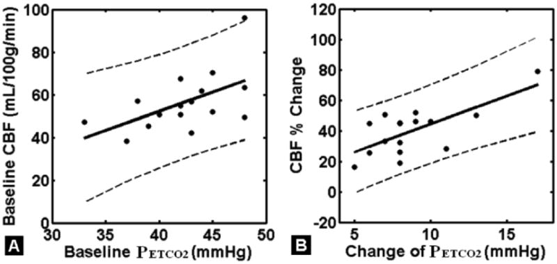
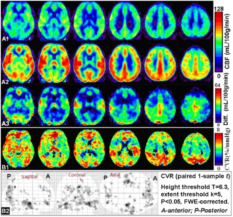
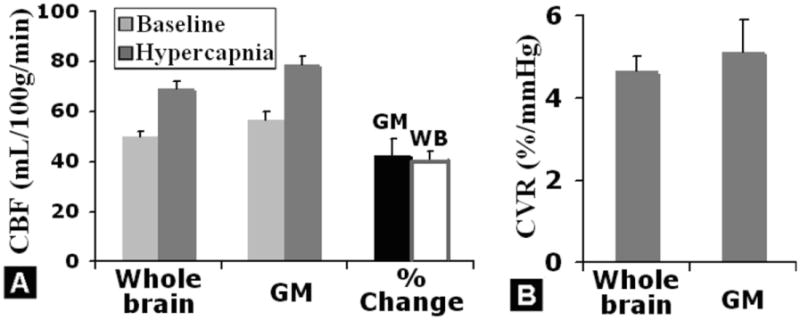
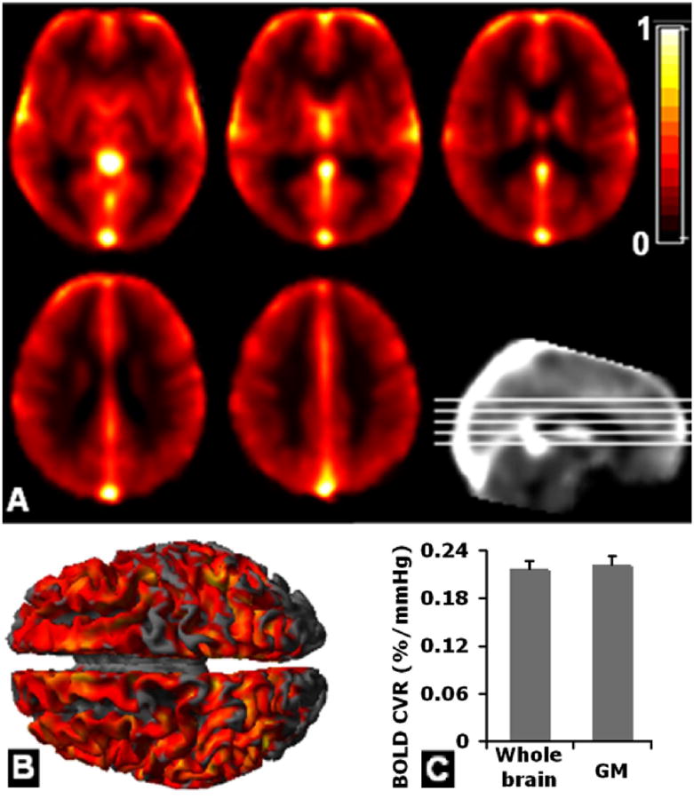
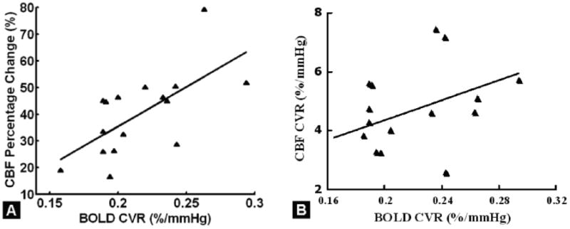
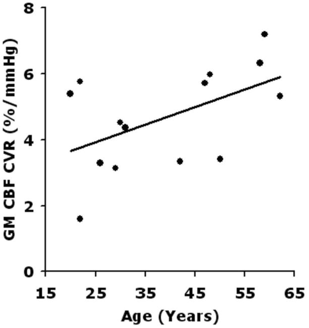
Results: The absolute CBF, BOLD and CVR measures were consistent with literature values. CBF derived CVR was 5.11±0.87%/mmHg in gray matter (GM) and 4.64±0.37%/mmHg in parenchyma. BOLD CVR was 0.23±0.04%/mmHg and 0.22±0.04%/mmHg for GM and parenchyma respectively. The most significant correlations between BOLD and CBF-based CVRs were also in GM structures, with greater vascular response in occipital cortex than in frontal and parietal lobes (6.8%/mmHg versus 4.5%/mmHg, 50% greater). Parenchymal BOLD CVR correlated significantly with the fractional change in CBF in response to hypercapnia (r=0.61, P=0.01), suggesting the BOLD response to be significantly flow driven. GM CBF decreased with age in room air (-5.58mL/100g/min per decade for GM; r=-0.51, P=0.05), but there was no association of CBF with age during hypercapnia. A trend toward increased pCASL CVR with age was observed, scaling as 0.64%/mmHg per decade for GM.
Conclusion: Consistent with previously reported CVR values, our results suggest that BOLD and CBF CVR techniques are complementary to each other in evaluating neuronal and vascular underpinning of hemodynamic processes.
Keywords: Arterial spin labeling; BOLD; Cerebral blood flow; Cerebrovascular reactivity; Hypercapnia-based fMRI calibration; Neurovascular coupling.
Copyright © 2015 Elsevier Inc. All rights reserved.
Figures


References
-
- Fülesdi B, Limburg M, Bereczki D. Cerebrovascular reactivity and reserve capacity in type II diabetes mellitus. J Diabetes Complications. 1999;13:191–199. - PubMed
-
- Spano VR, Mandell DM, Poublanc J, Sam K, Battisti-Charbonney A, Pucci O, et al. CO2 blood oxygen level-dependent MR mapping of cerebrovascular reserve in a clinical population: safety, tolerability, and technical feasibility. Radiology. 2013 Feb;266:592–8. - PubMed
-
- Yonas H, Smith HA, Durham SR, Pentheny SL, Johnson DW. Increased stroke risk predicted by compromised cerebral blood flow reactivity. J Neurosurg. 1993;79:483–489. - PubMed
-
- Rostrup E, Larsson HB, Toft PB, Garde K, Thomsen C, Ring P, et al. Functional MRI of CO2 induced increase in cerebral perfusion. NMR Biomed. 1994;7:29–34. - PubMed
-
- Rostrup E, Law I, Blinkenberg M, Larsson HBW, Born AP, Hom S, et al. Regional differences in the CBF and BOLD responses to hypercapnia: A combined PET and fMRI study. Neuroimage. 2000;11:87–97. - PubMed
Publication types
MeSH terms
Substances
Grants and funding
LinkOut - more resources
Full Text Sources
Other Literature Sources
Medical
