Review
. 2015 Apr;1342(1):44-52.
doi: 10.1111/nyas.12690. Epub 2015 Feb 23.

The pathogenesis of pediatric cerebral malaria: eye exams, autopsies, and neuroimaging

Affiliations
Review

The pathogenesis of pediatric cerebral malaria: eye exams, autopsies, and neuroimaging

Terrie E Taylor et al. Ann N Y Acad Sci. 2015 Apr.

Abstract

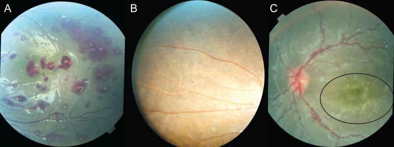
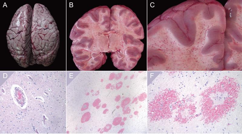
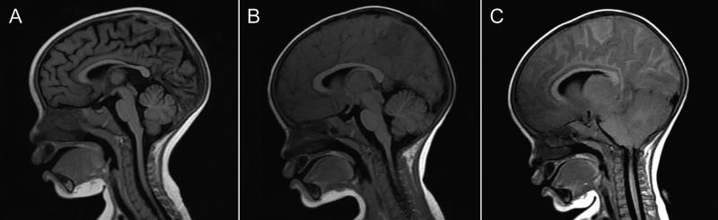
Several advances in our understanding of pediatric cerebral malaria (CM) have been made over the past 25 years. Accurate clinical diagnosis is enhanced by the identification of a characteristic retinopathy, visible by direct or indirect ophthalmoscopy, the retinal changes (retinal whitening, vessel color changes, white-centered hemorrhages) being consistently associated with intracerebral sequestration of parasites in autopsy studies. Autopsies have yielded information at tissue levels in fatal CM, but new insights into critical pathogenetic processes have emerged from neuroimaging studies, which, unlike autopsy-based studies, permit serial observations over time and allow comparisons between fatal cases and survivors. Brain swelling has emerged as the major risk factor for death, and, among survivors, brain volume diminishes spontaneously over 24-48 hours. Studies of life-threatening and fatal malaria are suggesting new approaches to identifying and caring for those at highest risk; potential adjuvants should be evaluated and implemented where they are most needed.

Keywords: African children; brain swelling; cerebral malaria; pathogenesis; sequestration.


Figures

Figure 1
Figure 1
Features of malarial retinopathy. Fundus photographs from pediatric patients in Malawi illustrate the 3 components of malarial retinopathy. (A)White centered hemorrhages. (B) Vessel color changes are evident in the branching veins in the center of this image. (C) Peri-macular whitening (shown within the circle) is distinct from the reflections created by the fundus camera
Figure 2
Figure 2
Postmortem findings (CM2). The gross and microscopic findings associated with the histologic subtype CM2 are shown here. (A) A slate gray appearance of the surface of the brain at autopsy. The flattened gyri and narrowed sulci are consistent with the MRI findings of increased brain volume in fatal pediatric CM. (B and C) A coronal section of the brain at the time of autopsy shows the characteristic distribution of hemorrhages in the white matter of the cerebral cortex. (D) A post-capillary venule with parasitized red cells surrounding a central column of unparasitized red cells. This appearance is also seen in CM1 patients. (E and F). Low and higher power images of ring hemorrhages in cortical white matter. Thrombi are present in the center of each vessel; extravasated erythrocytes are unparasitized.
Figure 3
Figure 3
MRI features in a fatal case of pediatric CM. These T2 images from the 0.35T MRI in Malawi illustrate the natural history of increasing brain volume in children with fatal CM. (A) MRI from a recovered patient, showing rounded gyri, in black, cerebrospinal fluid in the sulci and surrounding the brainstem. (B and C). These two studies were carried out on the same patient, 24 h apart. Flattened gyri and narrowed sulci are evident in both, and encroachment of the cerebellar tonsils on the brainstem, indicating herniation, is seen in C, 4 hours prior to death.
Figure 4
Figure 4
Identifying high-risk pediatric malaria patients. (A and B) In a group of children in a malaria-endemic region of sub-Saharan Africa, a proportion will be uninfected (A, pink), while others will have asymptomatic parasitemia (B, light purple). Some of these children will develop uncomplicated malaria (B, darker purple) and a few will progress to severe and complicated malaria (B, black). (C) Children meeting the standard clinical case definition of cerebral malaria are shown in (black). (D) Adding ocular funduscopic findings enhances the specificity of the diagnosis: retinopathy-negative patients are shown in blue, and the retinopathy-positive patients are yellow and red. (E) Only patients with retinopathy-positive CM, and among that group, the children at highest risk of a fatal outcome are those with brain swelling, shown in red in panels D, E, and F. Fatal cases are indicated with a cross.

Similar articles

Cited by

References

    1. Dondorp A, Nosten F, Stepniewska K, et al. Artesunate versus quinine for treatment of severe falciparum malaria: a randomised trial. Lancet. 2005;366:717–725. - PubMed
    1. von Seidlein L, Olaosebikan R, Hendriksen IC, et al. Clin Infect Dis. Vol. 54. United States: 2012. Predicting the clinical outcome of severe falciparum malaria in african children: findings from a large randomized trial. pp. 1080–1090. - PMC - PubMed
    1. Warrell DA, Looareesuwan S, Warrell MJ, et al. Dexamethasone proves deleterious in cerebral malaria. A double-blind trial in 100 comatose patients. N Engl J Med. 1982;306:313–319. - PubMed
    1. Molyneux ME, Taylor TE, Wirima JJ, et al. Clinical features and prognostic indicators in paediatric cerebral malaria: a study of 131 comatose Malawian children. Q J Med. 1989;71:441–459. - PubMed
    1. Newton CR, Chokwe T, Schellenberg JA, et al. Coma scales for children with severe falciparum malaria. Trans R Soc Trop Med Hyg. 1997;91:161–165. - PubMed