Review
. 2015 Mar;36(3):179-88.
doi: 10.1016/j.it.2015.02.001. Epub 2015 Feb 20.

RNA degradation in antiviral immunity and autoimmunity

Affiliations
Review

RNA degradation in antiviral immunity and autoimmunity

Rachel E Rigby et al. Trends Immunol. 2015 Mar.

Abstract

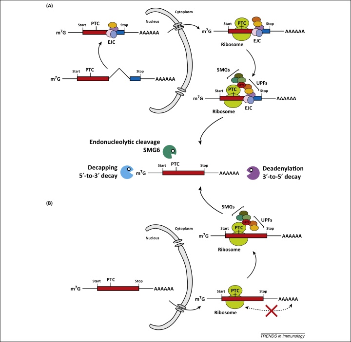
Post-transcriptional control determines the fate of cellular RNA molecules. Nonsense-mediated decay (NMD) provides quality control of mRNA, targeting faulty cellular transcripts for degradation by multiple nucleases including the RNA exosome. Recent findings have revealed a role for NMD in targeting viral RNA molecules, thereby restricting virus infection. Interestingly, NMD is also linked to immune responses at another level: mutations affecting the NMD or RNA exosome machineries cause chronic activation of defence programmes, resulting in autoimmune phenotypes. Here we place these observations in the context of other links between innate antiviral immunity and type I interferon mediated disease and examine two models: one in which expression or function of pathogen sensors is perturbed and one wherein host-derived RNA molecules with a propensity to activate such sensors accumulate.

Keywords: Aicardi-Goutières syndrome; MDA5; RNA exosome; nonsense-mediated decay; pattern-recognition receptor; type I interferon.


Figures

Figure 1
Figure 1
Nonsense-mediated decay (NMD). NMD recognises premature translation termination codons (PTCs) and this requires translation. The mechanism of PTC definition differs between species and individual transcripts. Two major pathways are shown here. (A) One important mechanism of PTC recognition relies on a protein complex called the exon-junction complex (EJC) . EJCs are deposited on mRNAs during splicing and mark exon-exon junctions. EJCs are transported together with the mRNA into the cytoplasm and are removed from the mRNA during translation [82–84]. Importantly, most mRNAs contain the stop codon in their last exon; therefore, no EJCs are left on the mRNA when translation termination occurs. However, if mRNAs have a PTC upstream of the last exon, one or multiple EJCs remain on the mRNA at the moment when translation terminates. This constellation is recognised by a number of proteins including the essential NMD effectors UPF1, UPF2, and UPF3/3X. UPF1 (red) interacts with proteins involved in translation termination, while UPF2 (orange) and UPF3/3X (yellow) associate with the EJC. If an EJC is present downstream from a terminating ribosome, the UPF proteins interact to form a complex, SMG proteins (olive) are recruited, and degradation of the mRNA is initiated (middle panel) [5–8]. (B) EJCs are not always required for NMD [5–8]. Efficient translation termination requires interactions between proteins bound to the mRNA poly(A) tail and release factors, which associate with ribosomes at stop codons. If termination occurs at a PTC, the distance to the 3′-end and poly(A) tail may be too large to accommodate this interaction (red cross) [25,85–87]. This in turn is presumed to result in delayed release of the ribosome from the mRNA, allowing for assembly of UPF proteins and recruitment of SMGs independently of an EJC.
Figure 2
Figure 2
Nucleic acid detection by pattern recognition receptors. Virus infection delivers nucleic acids into infected cells. (Left) DNA is detected in the cytoplasm by cyclic GMP-AMP synthase (cGAS), which then uses ATP and GTP to generate cyclic GMP-AMP (cGAMP). This second messenger subsequently binds to the ER-associated protein STING which in turn activates TBK1. This kinase then phosphorylates the transcription factor IRF3, which forms a dimer and translocates to the cell nucleus to induce type I IFN gene transcription. Aside from cGAS, additional proteins including IFI16, DDX41, MRE11, and DNA-PK may participate in cytosolic DNA recognition but are not shown here for simplicity. (Centre) Viral nucleic acids are also detected in endosomal compartments by TLR9 (DNA), TLR3 (dsRNA), and TLR7/8 (ssRNA). These PRRs signal through the adaptor proteins MyD88 or TRIF to activate the kinases IKKα, TBK1, and IKKɛ resulting in phosphorylation, dimerisation and translocation of IRF3 or IRF7. (Right) RNA in the cytoplasm of infected cells is recognised by RIG-I and MDA5, which interact with the mitochondrial protein MAVS to trigger TBK1/IKKɛ. Please note that a variety of other proteins are involved in signal transduction that are not shown here for clarity.

References

    1. Garcia D. Nonsense-mediated decay serves as a general viral restriction mechanism in plants. Cell Host Microbe. 2014;16:391–402. - PMC - PubMed
    1. Balistreri G. The host nonsense-mediated mRNA decay pathway restricts Mammalian RNA virus replication. Cell Host Microbe. 2014;16:403–411. - PubMed
    1. Gloggnitzer J. Nonsense-mediated mRNA decay modulates immune receptor levels to regulate plant antibacterial defense. Cell Host Microbe. 2014;16:376–390. - PubMed
    1. Eckard S.C. The SKIV2L RNA exosome limits activation of the RIG-I-like receptors. Nat. Immunol. 2014;15:839–845. - PMC - PubMed
    1. Behm-Ansmant I. mRNA quality control: an ancient machinery recognizes and degrades mRNAs with nonsense codons. FEBS Lett. 2007;581:2845–2853. - PubMed

Publication types

MeSH terms

Supplementary concepts

LinkOut - more resources