

Case Reports
doi: 10.2147/OTT.S68819.
eCollection 2015.
The imaging features of metanephric adenoma: a case report and review of literature
Affiliations
- PMID: 25709478
- PMCID: PMC4335625
- DOI: 10.2147/OTT.S68819
Item in Clipboard
Case Reports
The imaging features of metanephric adenoma: a case report and review of literature
Onco Targets Ther.
.
Abstract
Metanephric adenoma (MA) is a rare epithelial tumor of the kidney with a characteristic histology. To date, the imaging features of the tumor have not been clearly described. Until now, MA was considered to be benign, but the majority of MA cases underwent nephrectomy. Here, we report a case of MA confirmed by surgical pathology, and we will analyze the ultrasound and computed tomography findings. The radiological features of MA are presented along with a brief review of the pertinent literature to deepen the understanding of MA's imaging features.
Keywords: X-ray computed tomography; histology; metanephric adenoma; ultrasound.
Figures
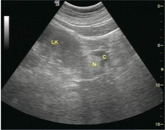
Longitudinal sonogram of the left kidney. Notes: Longitudinal sonogram shows a 3.2 cm ×4.5 cm liquid dark area (C) in the lower pole of the left kidney (LK). An irregular 2.4 cm ×2.1 cm, slightly hyperechoic mass (N) is visible in the liquid dark area without acoustical shadowing, which was extensively connected to the left kidney.
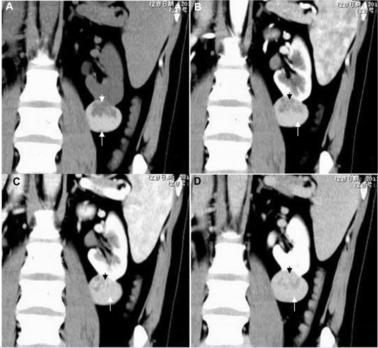
CT images of the left renal tumor. Notes: (A) Unenhanced CT scan shows a well-defined, high-density mass measuring 3.7 cm ×3.3 cm (long arrow) in the lower pole of the left kidney, with an irregularly shaped lower attenuation area (short arrow) in the mass. (B–D) Enhanced CT scan shows that the tumor appears to have no enhancement in the peripheral portion (white arrow), and progressive enhancement in the central portion (black arrow). Abbreviation: CT, computed tomography.
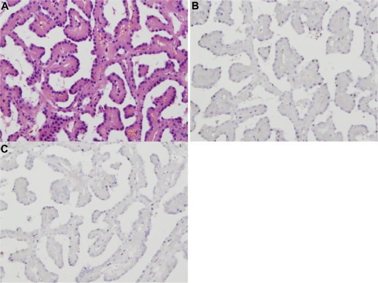
Microscopic appearance of MA. Notes: (A) A histological specimen shows the homogeneous round or ovoid tumor cells mainly composed of tiny tubules and papillae, accompanied by very scanty stroma (HE ×200); (B and C) the tumor cells are negative for WT1 and CD57 (SP ×200), respectively. Abbreviations: MA, metanephric adenoma; HE, hematoxylin and eosin; SP, streptavidin-peroxidase.
Similar articles
-
Metanephric Adenoma in the Pediatric Population: Diagnostic Challenges and Follow-up.Urology. 2018 Oct;120:211-215. doi: 10.1016/j.urology.2018.06.042. Epub 2018 Jul 10. Urology. 2018. PMID: 30006267 Review.
-
Metanephric adenoma of the kidney: a case report.Pediatr Radiol. 1999 Feb;29(2):100-3. doi: 10.1007/s002470050550. Pediatr Radiol. 1999. PMID: 9933328
-
Metanephric adenoma in an 8-year-old child: case report and review of the literature.J Pediatr Surg. 2005 May;40(5):e25-8. doi: 10.1016/j.jpedsurg.2005.02.019. J Pediatr Surg. 2005. PMID: 15937802 Review.
-
Metanephric adenoma: A report of two cases and review of the literature.Mol Clin Oncol. 2013 Nov;1(6):1087-1089. doi: 10.3892/mco.2013.184. Epub 2013 Sep 13. Mol Clin Oncol. 2013. PMID: 24649298 Free PMC article.
-
The findings of CT and MRI in patients with metanephric adenoma.Diagn Pathol. 2016 Oct 27;11(1):104. doi: 10.1186/s13000-016-0535-x. Diagn Pathol. 2016. PMID: 27784295 Free PMC article.
Cited by
-
Case Report: MRI, CEUS, and CT Imaging Features of Metanephric Adenoma with Histopathological Correlation and Literature Review.Diagnostics (Basel). 2022 Aug 26;12(9):2071. doi: 10.3390/diagnostics12092071. Diagnostics (Basel). 2022. PMID: 36140473 Free PMC article.
-
Renal Tumors of Childhood: Radiologic-Pathologic Correlation Part 2. The 2nd Decade: From the Radiologic Pathology Archives.Radiographics. 2017 Sep-Oct;37(5):1538-1558. doi: 10.1148/rg.2017160189. Radiographics. 2017. PMID: 28898190 Free PMC article. Review.
-
Experience of diagnosis and management of metanephric adenoma: retrospectively analysis of 10 cases and a literature review.Transl Androl Urol. 2020 Aug;9(4):1661-1669. doi: 10.21037/tau-19-912. Transl Androl Urol. 2020. PMID: 32944527 Free PMC article.
-
Metanephric Adenoma: A Case Report of a Rare Epithelial Renal Tumor.Cureus. 2024 Apr 18;16(4):e58545. doi: 10.7759/cureus.58545. eCollection 2024 Apr. Cureus. 2024. PMID: 38957819 Free PMC article.
References
-
- Brisigotti M, Cozzutto C, Fabbretti G, et al. Metanephric adenoma. Histol Histopathol. 1992;7(4):689–692. - PubMed
-
- Davis CJ, Jr, Barton JH, Sesterhenn IA, et al. Metanephric adenoma. Clinicopathological study of fifty patients. Am J Surg Pathol. 1995;19(10):1101–1114. - PubMed
-
- Jemal A, Bray F, Center MM, et al. Global cancer statistics. CA Cancer J Clin. 2011;61(2):69–90. - PubMed
Publication types
LinkOut - more resources
Full Text Sources
