

The Added Value of a Single-photon Emission Computed Tomography-Computed Tomography in Sentinel Lymph Node Mapping in Patients with Breast Cancer and Malignant Melanoma
- PMID: 25709544
- PMCID: PMC4337007
- DOI: 10.4103/1450-1147.150543
The Added Value of a Single-photon Emission Computed Tomography-Computed Tomography in Sentinel Lymph Node Mapping in Patients with Breast Cancer and Malignant Melanoma
Abstract
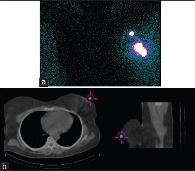
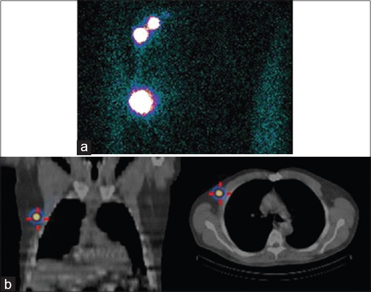
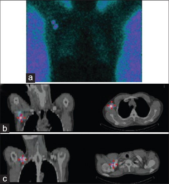
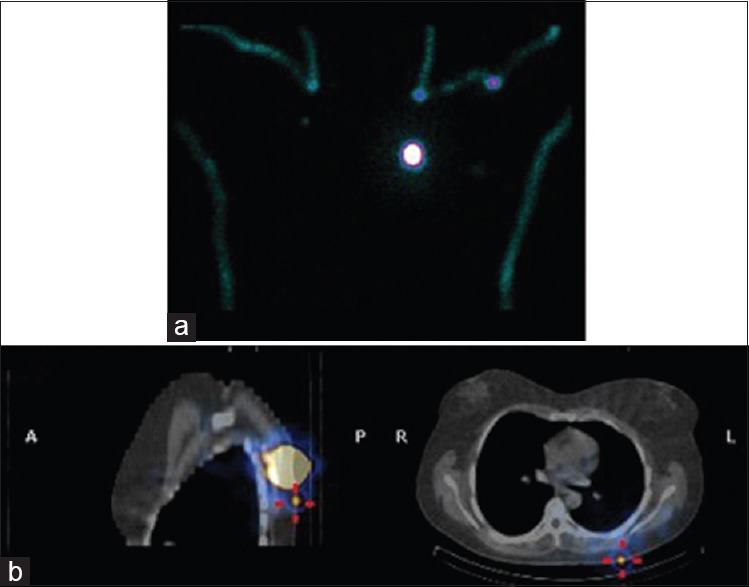
Single-photon emission computed tomography-computed tomography (SPECT-CT) allows for physiological and anatomical co-registration in sentinel lymph node (SLN) mapping and offers additional benefits over conventional planar imaging. However, the clinical relevance when considering added costs and radiation burden of these reported benefits remains somewhat uncertain. This study aimed to evaluate the possible added value of SPECT-CT and intra-operative gamma-probe use over planar imaging alone in the South African setting. 80 patients with breast cancer or malignant melanoma underwent both planar and SPECT-CT imaging for SLN mapping. We assessed and compared the number of nodes detected on each study, false positive and negative findings, changes in surgical approach and or patient management. In all cases where a sentinel node was identified, SPECT-CT was more accurate anatomically. There was a significant change in surgical approach in 30 cases - breast cancer (n = 13; P 0.001) and malignant melanoma (n = 17; P 0.0002). In 4 cases a node not identified on planar imaging was seen on SPECT-CT. In 16 cases additional echelon nodes were identified. False positives were excluded by SPECT-CT in 12 cases. The addition of SPECT-CT and use of intra-operative gamma-probe to planar imaging offers important benefits in patients who present with breast cancer and melanoma. These benefits include increased nodal detection, elimination of false positives and negatives and improved anatomical localization that ultimately aids and expedites surgical management. This has been demonstrated in the context of industrialized country previously and has now also been confirmed in the setting of a emerging-market nation.
Keywords: Breat cancer; melanoma; sentinel lymph node; single-photon emission computed tomography-computed tomography.
Conflict of interest statement
Figures
Similar articles
-
Utility of single-photon emission tomography/computed tomography for sentinel lymph node localization in breast cancer patients.Nucl Med Commun. 2017 Jun;38(6):493-499. doi: 10.1097/MNM.0000000000000676. Nucl Med Commun. 2017. PMID: 28430742
-
Clinical Value of Single-photon Emission Computed Tomography Combined With Computed Tomography for Sentinel Lymph Node Identification in Melanoma.Ann Plast Surg. 2019 Apr;82(4S Suppl 3):S192-S194. doi: 10.1097/SAP.0000000000001895. Ann Plast Surg. 2019. PMID: 30855387
-
Evaluation of the diagnostic value of preoperative sentinel lymph node (SLN) imaging in penile carcinoma patients without palpable inguinal lymph nodes via single photon emission computed tomography/computed tomography (SPECT/CT) as compared to planar scintigraphy.Urol Oncol. 2018 Mar;36(3):92.e17-92.e24. doi: 10.1016/j.urolonc.2017.11.012. Epub 2017 Dec 14. Urol Oncol. 2018. PMID: 29249274
-
Single-photon emission computed tomography/computed tomographyfor sentinel node mapping in breast cancer.Semin Nucl Med. 2007 Jan;37(1):29-33. doi: 10.1053/j.semnuclmed.2006.08.001. Semin Nucl Med. 2007. PMID: 17161037 Review.
-
Contribution of SPECT/CT imaging to radioguided sentinel lymph node biopsy in breast cancer, melanoma, and other solid cancers: from "open and see" to "see and open".Q J Nucl Med Mol Imaging. 2014 Jun;58(2):127-39. Q J Nucl Med Mol Imaging. 2014. PMID: 24835289 Review.
Cited by
-
Multi-slice SPECT/CT vs. lymphoscintigraphy and intraoperative gamma ray probe for sentinel node mapping in HNSCC.Eur Arch Otorhinolaryngol. 2017 Mar;274(3):1633-1642. doi: 10.1007/s00405-016-4379-5. Epub 2016 Nov 11. Eur Arch Otorhinolaryngol. 2017. PMID: 27837418
-
The Usefulness of SPECT/CT in Sentinel Node Mapping of Early Stage Breast Cancer Patients Showing Negative or Equivocal Findings on Planar Scintigraphy.Asia Ocean J Nucl Med Biol. 2018 Spring;6(2):80-89. doi: 10.22038/aojnmb.2018.10720. Asia Ocean J Nucl Med Biol. 2018. PMID: 29998140 Free PMC article.
-
Comparison of 99mTc-Labeled Colloid SPECT/CT and Planar Lymphoscintigraphy in Sentinel Lymph Node Detection in Patients with Melanoma: A Meta-Analysis.J Clin Med. 2020 Jun 2;9(6):1680. doi: 10.3390/jcm9061680. J Clin Med. 2020. PMID: 32498217 Free PMC article. Review.
References
-
- Davis PG, Serpell JW, Kelly JW, Paul E. Axillary lymph node dissection for malignant melanoma. ANZ J Surg. 2011;81:462–6. - PubMed
-
- Buscombe J, Paganelli G, Burak ZE, Waddington W, Maublant J, Prats E, et al. Sentinel node in breast cancer procedural guidelines. Eur J Nucl Med Mol Imaging. 2007;34:2154–9. - PubMed
-
- Herbst M. In: Cancer Association of South Africa: Fact Sheet on Solar Radiation and Skin Cancer. van Rensburg SJ, editor. Johannesburg, South Africa: CANSA; 2013. pp. 1–19.
-
- van der Ploeg IM, Valdés Olmos RA, Nieweg OE, Rutgers EJ, Kroon BB, Hoefnagel CA. The additional value of SPECT/CT in lymphatic mapping in breast cancer and melanoma. J Nucl Med. 2007;48:1756–60. - PubMed
-
- Center M, Siegel R, Jemal A, editors. Global Cancer Facts and Figures. 2nd ed. Atlanta, Georgia: American Cancer Society; 2008.
LinkOut - more resources
Full Text Sources
Other Literature Sources