Review
. 2015 Feb 9:9:23.
doi: 10.3389/fnins.2015.00023. eCollection 2015.

Methylglyoxal, the dark side of glycolysis

Affiliations
Review

Methylglyoxal, the dark side of glycolysis

Igor Allaman et al. Front Neurosci. .

Abstract

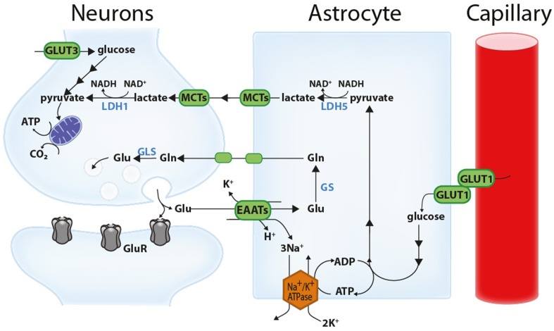
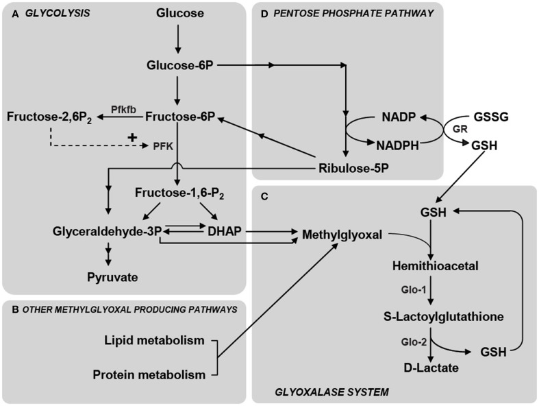
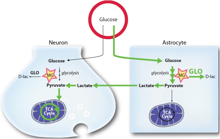
Glucose is the main energy substrate for the brain. There is now extensive evidence indicating that the metabolic profile of neural cells with regard to glucose utilization and glycolysis rate is not homogenous, with a marked propensity for glycolytic glucose processing in astrocytes compared to neurons. Methylglyoxal, a highly reactive dicarbonyl compound, is inevitably formed as a by-product of glycolysis. Methylglyoxal is a major cell-permeant precursor of advanced glycation end-products (AGEs), which are associated with several pathologies including diabetes, aging and neurodegenerative diseases. In normal situations, cells are protected against methylglyoxal toxicity by different mechanisms and in particular the glyoxalase system, which represents the most important pathway for the detoxification of methylglyoxal. While the neurotoxic effects of methylglyoxal and AGEs are well characterized, our understanding the glyoxalase system in the brain is more scattered. Considering the high energy requirements (i.e., glucose) of the brain, one should expect that the cerebral glyoxalase system is adequately fitted to handle methylglyoxal toxicity. This review focuses on our actual knowledge on the cellular aspects of the glyoxalase system in brain cells, in particular with regard to its activity in astrocytes and neurons. A main emerging concept is that these two neural cell types have different and energetically adapted glyoxalase defense mechanisms which may serve as protective mechanism against methylglyoxal-induced cellular damage.

Keywords: advanced-glycation end-products (AGEs); astrocyte; glutathione; methylglyoxal; neuron; triosephosphate.


Figures

Figure 1
Figure 1
Schematic representation of the astrocyte-neuron lactate shuttle (ANLS). Glutamate (Glu) released at the synapse activates glutamatergic receptors (GluR) and is associated with important energy expenditures in neuronal compartments. A large proportion of the glutamate released at the synapse is taken up by astrocytes via excitatory amino acid transporters (EAATs, more specifically GLT-1 and GLAST) together with 3 Na+ ions. This Na+ is extruded by the action of the Na+/K+ ATPase, consuming ATP. This triggers non oxidative glucose utilization in astrocytes and glucose uptake from the circulation through the glucose transporter GLUT1 expressed by both capillary endothelial cells and astrocytes. Glycolytically-derived pyruvate is converted to lactate by lactate dehydrogenase 5 (LDH5; mainly expressed in astrocytes) and shuttled to neurons through monocarboxylate transporters (mainly MCT1 and MCT4 in astrocytes and MCT2 in neurons). In neurons, this lactate can be used as an energy substrate following its conversion to pyruvate by LDH1 (mainly expressed in neurons). Neurons can also take up glucose via the neuronal glucose transporter 3 (GLUT3). Concomitantly, astrocytes participate in the recycling of synaptic glutamate via the glutamate-glutamine cycle. Following its uptake by astrocytes, glutamate is converted to glutamine (Gln) by the action of glutamine synthetase (GS) and shuttled to neurons where it is converted back to glutamate by glutaminases (GLS). Reproduced with permission from Bélanger et al. (2011a).
Figure 2
Figure 2
Schematic representation of the main metabolic pathways involved in MG production and elimination. (A) MG is formed mainly by the fragmentation of the glycolytic intermediates glyceraldehyde-3-phosphate (Glyceraldehyde-3P) and dihydroxyacetone phosphate (DHAP), but also from the metabolism of lipids and proteins (B). Phosphofructokinase-1 (PFK) is the rate-limiting step of glycolysis and thus constitutes an important regulatory site, one of its most potent allosteric activators being Fructose-2,6-bisphosphate (Fructose-2,6-P2). Fructose-2,6-P2 levels are controlled by the enzyme 6-phosphofructose-2-kinase/fructose-2,6-bisphosphatase (Pfkfb) which is most abundantly expressed in astrocytes compared to neurons (Herrero-Mendez et al., 2009). (C) MG is detoxified principally via the glyoxalase system which consists of the enzymes Glyoxalase-1 (Glo-1) and Glyoxalase-2 (Glo-2). The first step in MG detoxification requires its spontaneous reaction with reduced glutathione (GSH) to form a hemithioacetal which is used as a substrate by Glo-1 to form S-Lactoylglutathione. Glo-2 then catalyzes the transformation of S-Lactoylglutathione into D-Lactate, recycling GSH in the process. (D) The pentose phosphate pathway is linked to MG detoxification via the formation of NADPH which is required for the recycling of GSH from its oxidized form (GSSG) via the action of glutathione reductase (GR). Reproduced with permission from Bélanger et al. (2011b).
Figure 3
Figure 3
Schematic representation of the proposed mechanism by which higher glycolytic rates in astrocytes may provide a mechanism limiting MG toxicity in neurons. Glycolysis in both astrocytes and neurons leads to the production of the toxic MG by-product. MG is detoxified by both cell types through the glyoxalase system (GLO), producing D-lactate (D-lac). In normal and stimulated conditions (ANLS) glucose utilization and its processing through the glycolysis is tilted toward astrocytes, which release L-lactate (lactate) that can be used by neurons as a mitochondrial energy substrate. Due to the high activity of the MG-detoxifying glyoxalase system in astrocytes, these cells are well equipped to handle MG accumulation and toxicity. Because they display lower glyoxalase system activity, neurons benefit from the glycolytic processing of glucose in astrocytes since they are spared from: (1) MG accumulation and toxicity (2) alterations in their antioxidant status (through the mobilization of GSH by Glo-1), and (3) the burden of mounting an enzymatic system to process MG. See text for more details. Green arrows highlight the prevalent routes of glucose utilization in brain cells.

Similar articles

Cited by

References

    1. Ahmed N., Battah S., Karachalias N., Babaei-Jadidi R., Horanyi M., Baroti K., et al. . (2003a). Increased formation of methylglyoxal and protein glycation, oxidation and nitrosation in triosephosphate isomerase deficiency. Biochim. Biophys. Acta 1639, 121–132. 10.1016/j.bbadis.2003.08.002 - DOI - PubMed
    1. Ahmed N., Thornalley P. J., Dawczynski J., Franke S., Strobel J., Stein G., et al. . (2003b). Methylglyoxal-derived hydroimidazolone advanced glycation end-products of human lens proteins. Invest. Ophthalmol. Vis. Sci. 44, 5287–5292. 10.1167/iovs.03-0573 - DOI - PubMed
    1. Allaman I. (2009). Glial glycogen metabolism, in Encyclopedia of Neuroscience, ed Squire L. R. (Oxford: Academic Press; ), 811–818.
    1. Allaman I., Belanger M., Magistretti P. J. (2011). Astrocyte-neuron metabolic relationships: for better and for worse. Trends Neurosci. 34, 76–87. 10.1016/j.tins.2010.12.001 - DOI - PubMed
    1. Allaman I., Gavillet M., Belanger M., Laroche T., Viertl D., Lashuel H. A., et al. . (2010). Amyloid-beta aggregates cause alterations of astrocytic metabolic phenotype: impact on neuronal viability. J. Neurosci. 30, 3326–3338. 10.1523/JNEUROSCI.5098-09.2010 - DOI - PMC - PubMed