Review
. 2014 Dec 15:13:95-100.
doi: 10.1016/j.csbj.2014.12.004. eCollection 2015.

Tissue-nonspecific Alkaline Phosphatase Regulates Purinergic Transmission in the Central Nervous System During Development and Disease

Affiliations
Review

Tissue-nonspecific Alkaline Phosphatase Regulates Purinergic Transmission in the Central Nervous System During Development and Disease

Álvaro Sebastián-Serrano et al. Comput Struct Biotechnol J. .

Abstract

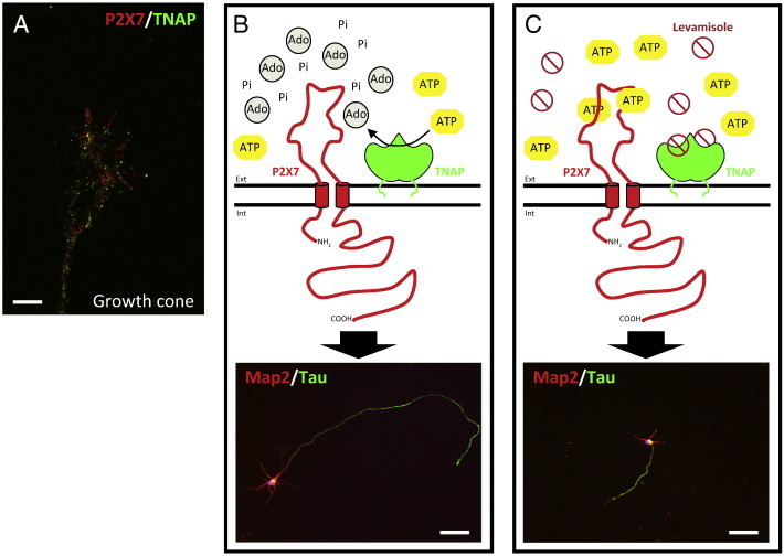
Tissue-nonspecific alkaline phosphatase (TNAP) is one of the four isozymes in humans and mice that have the capacity to hydrolyze phosphate groups from a wide spectrum of physiological substrates. Among these, TNAP degrades substrates implicated in neurotransmission. Transgenic mice lacking TNAP activity display the characteristic skeletal and dental phenotype of infantile hypophosphatasia, as well as spontaneous epileptic seizures and die around 10 days after birth. This physiopathology, linked to the expression pattern of TNAP in the central nervous system (CNS) during embryonic stages, suggests an important role for TNAP in neuronal development and synaptic function, situating it as a good target to be explored for the treatment of neurological diseases. In this review, we will focus mainly on the role that TNAP plays as an ectonucleotidase in CNS regulating the levels of extracellular ATP and consequently purinergic signaling.

Keywords: ATP; Development; Neurodegenerative diseases; P2X7R; Pyridoxal phosphate; TNAP.


Figures

Fig. 1
Fig. 1
Schematic representation of the axonal growth regulation by the coordinated action of TNAP and P2X7R. A) Immunofluorescence image of the axonal growth of hippocampal neurons fixed at 3 DIV and stained with antibodies against TNAP (green) and P2X7R (red). The image shows the presence of both proteins at the growth cone of hippocampal neurons. Scale bar: 20 μm. B) TNAP hydrolyzes the physiological agonist of P2X7R, ATP, in the proximal environment of the receptor, which negatively regulates the activation of this receptor favoring in this way the axonal growth. C) The pharmacological inhibition of TNAP by levamisole produces an increase of ATP in the proximal environment of P2X7R, event that favor the activation of the receptor and then decreasing the axonal growth. The immunofluorescence images of lower panels show hippocampal neurons (3 DIV) stained with antibodies against axonal molecular markers, Map2 (red) and Tau (green), under normal condition (B) or treated with TNAP antagonist, levamisole, inhibiting axonal growth (C). Scale bar, B and C: 50 μm. Ext: extracellular space. Int: intracellular space. Ado: adenosine. Pi: inorganic phosphate. (For interpretation of the references to color in this figure legend, the reader is referred to the web version of this article.)
Fig. 2
Fig. 2
Schematic representation illustrating the involvement of TNAP in the progression of Alzheimer's disease. First, tau protein reaches the interstitial space as a consequence of an initial undetermined injury or by its own vesicular release under physiological conditions. This tau protein is dephosphorylated by TNAP becoming an active ligand of muscarinic receptors. The activation of muscarinic receptors by tau has three main consequences; an increase of intracellular level of calcium and hyperphosphorylated tau, and an increase of TNAP levels. Subsequently of these effects, a positive feedback loop is generated in which the final consequence is cell death. With the rupture of the plasma membrane after the cell death, the intracellular contents are released to the interstitial space, increasing the extracellular levels of hyperphosphorylated tau. The NFTs (intracellular neurofibrillary tangles) suffer slow disassembly and degradation, allowing the proteins to reach distant brain regions that results in spreading of this neurodegenerative process.

References

    1. McComb R.B., Bowers G.N., Posen S. Plenum Press; New York: 1979. Alkaline phosphatase.
    1. Millan J.L. Alkaline phosphatases: structure, substrate specificity and functional relatedness to other members of a large superfamily of enzymes. Purinergic Signal. 2006;2:335–341. - PMC - PubMed
    1. al-Rashida M., Iqbal J. Therapeutic potentials of ecto-nucleoside triphosphate diphosphohydrolase, ecto-nucleotide pyrophosphatase/phosphodiesterase, ecto-5′-nucleotidase, and alkaline phosphatase inhibitors. Med Res Rev. 2014;34:703–743. - PubMed
    1. Harris H. The human alkaline phosphatases: what we know and what we don't know. Clin Chim Acta. 1990;186:133–150. - PubMed
    1. Van Hoof V.O., De Broe M.E. Interpretation and clinical significance of alkaline phosphatase isoenzyme patterns. Crit Rev Clin Lab Sci. 1994;31:197–293. - PubMed