

Congenital portosystemic shunt: our experience
- PMID: 25709849
- PMCID: PMC4332460
- DOI: 10.1155/2015/691618
Congenital portosystemic shunt: our experience
Abstract
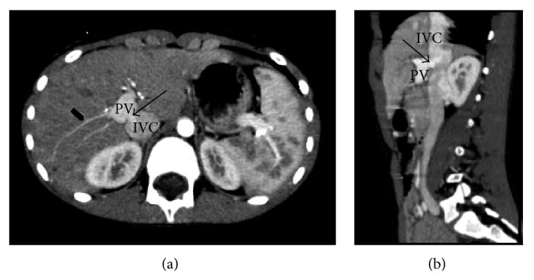
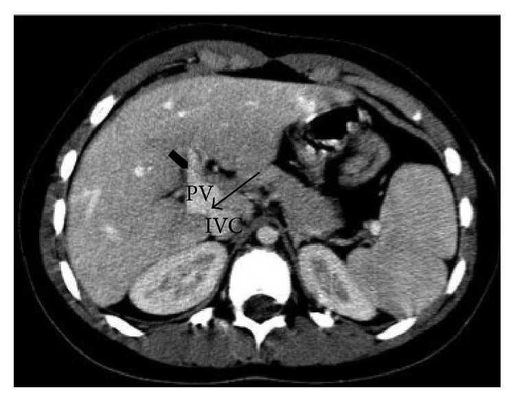
Introduction. Congenital portosystemic venous malformations are rare abnormalities in which the portal blood drains into a systemic vein and which are characterized by extreme clinical variability. Case Presentations. The authors present two case reports of a congenital extrahepatic portosystemic shunt (Type II). In the first patient, apparently nonspecific symptoms, such as headache and fatigue, proved to be secondary to hypoglycemic episodes related to the presence of a portosystemic shunt, later confirmed on imaging. During portal vein angiography, endovascular embolization of the portocaval fistula achieved occlusion of the anomalous venous tract. In the second patient, affected by Down's syndrome, the diagnosis of a portosystemic malformation was made by routine ultrasonography, performed to rule out concurrent congenital anomalies. Because of the absence of symptoms, we chose to observe this patient. Conclusions. These two case reports demonstrate the clinical heterogeneity of this malformation and the need for a multidisciplinary approach. As part of a proper workup, clinical evaluation must always be followed by radiographic diagnosis.
Figures
References
-
- Writters P., Maleaux G., George C., et al. Congenital veno-venous malformations of the liver: widely variable clinical presentations. Journal of Gastroenterology and Hepatology. 2008;23(8, part 2):e390–e394. - PubMed
LinkOut - more resources
Full Text Sources
Other Literature Sources
Research Materials
